Loading video…

 18 Concepts
 4.3K views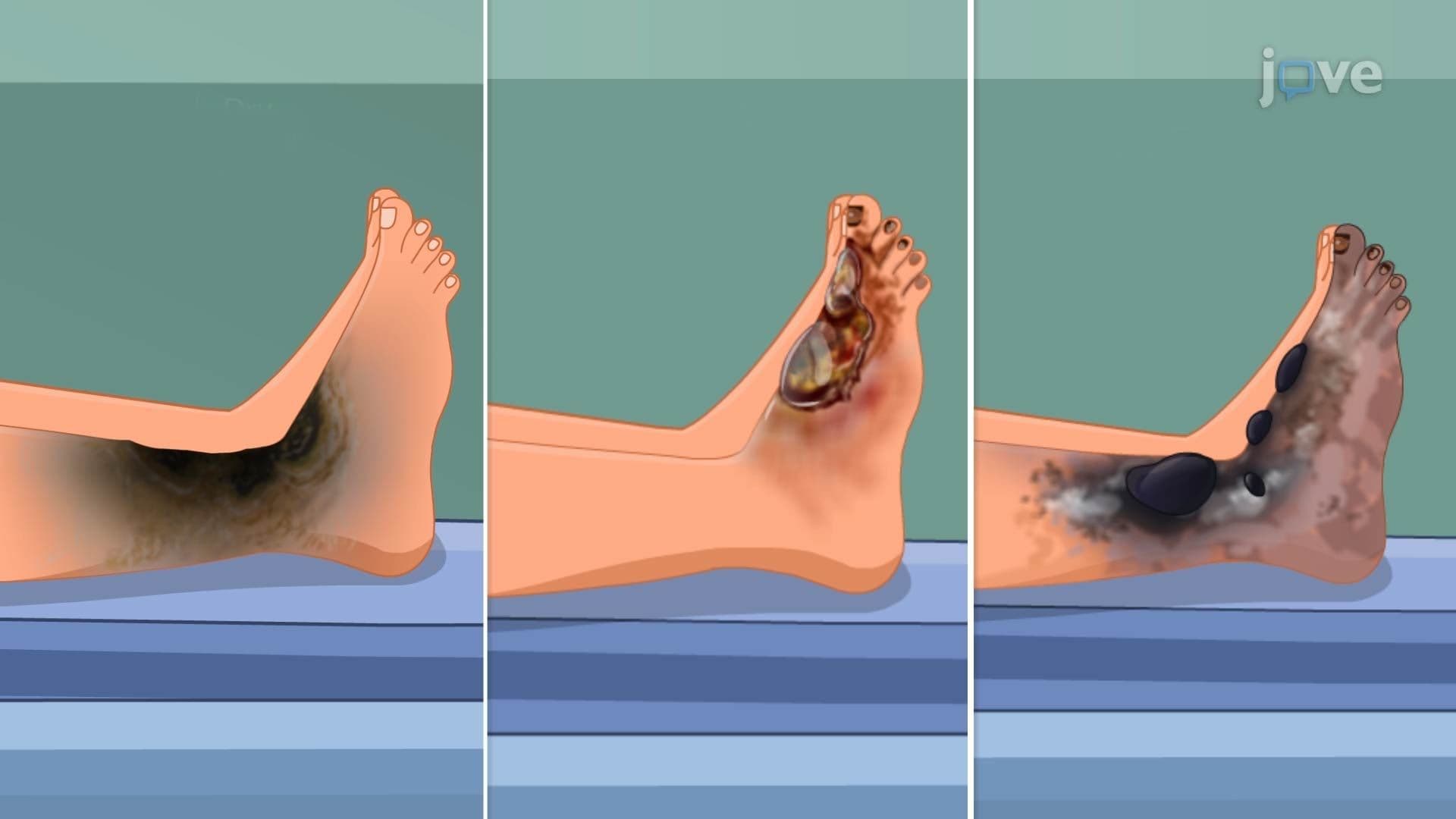

Video Summary: Necrosis in Cellular Injury Iv
When a cell can no longer survive an injury, it doesn't simply shut down, it ruptures, spills its contents, and triggers a full inflammatory response. That dramatic process is what Necrosis in Cellular Injury IV explores. In US hospital settings, necrotic tissue is commonly seen in patients with severe frostbite or diabetic foot ulcers. Understanding necrosis helps explain why damaged tissue looks and behaves so differently across the body. Watch the full video on JoVE Coach to master this concept with expert-led visuals and step-by-step explanations.
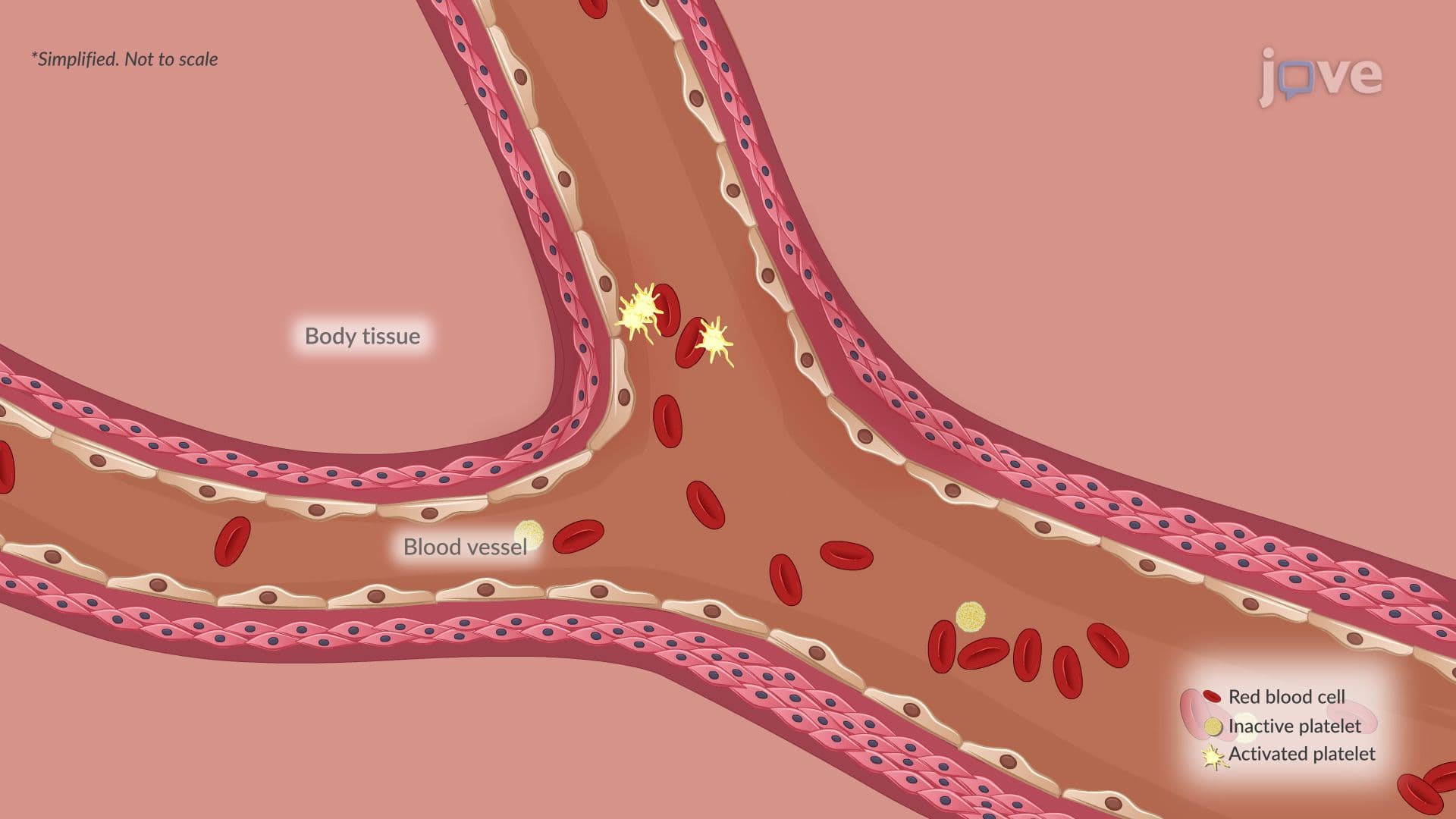
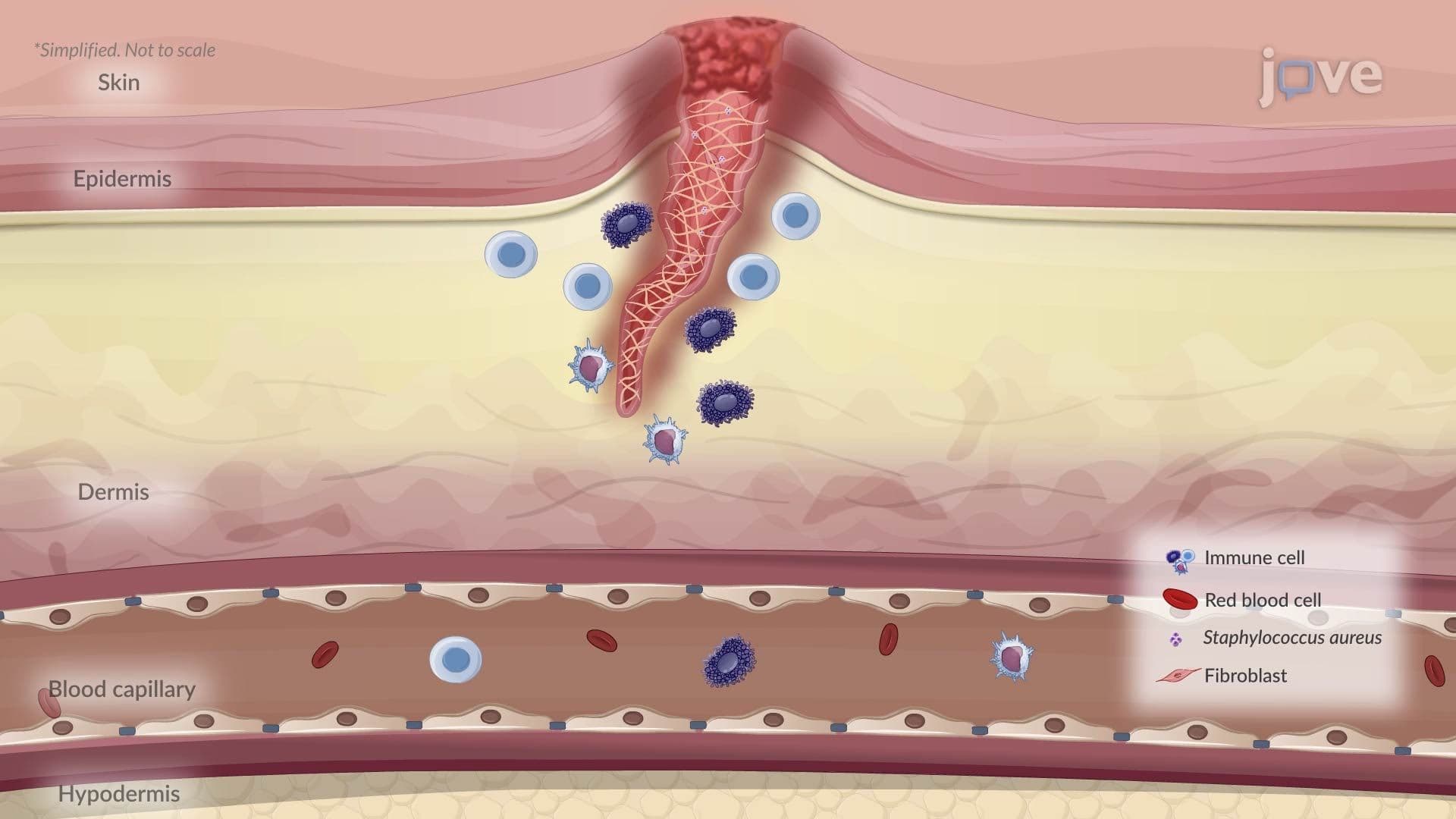
Necrosis represents one of the most consequential outcomes of severe cellular injury. Unlike apoptosis, the body's controlled, "programmed" method of cell removal, necrosis is unplanned, destructive, and provokes a strong immune response. It occurs when a cell experiences injury so severe that it cannot maintain basic functions. Common triggers include ischemia (restricted blood flow, as seen in heart attacks), bacterial toxins (such as those from *Staphylococcus aureus* infections), and physical trauma. Understanding necrosis is essential for any student studying disease mechanisms, because it helps answer the fundamental question: *How does the body respond to cellular injury?*
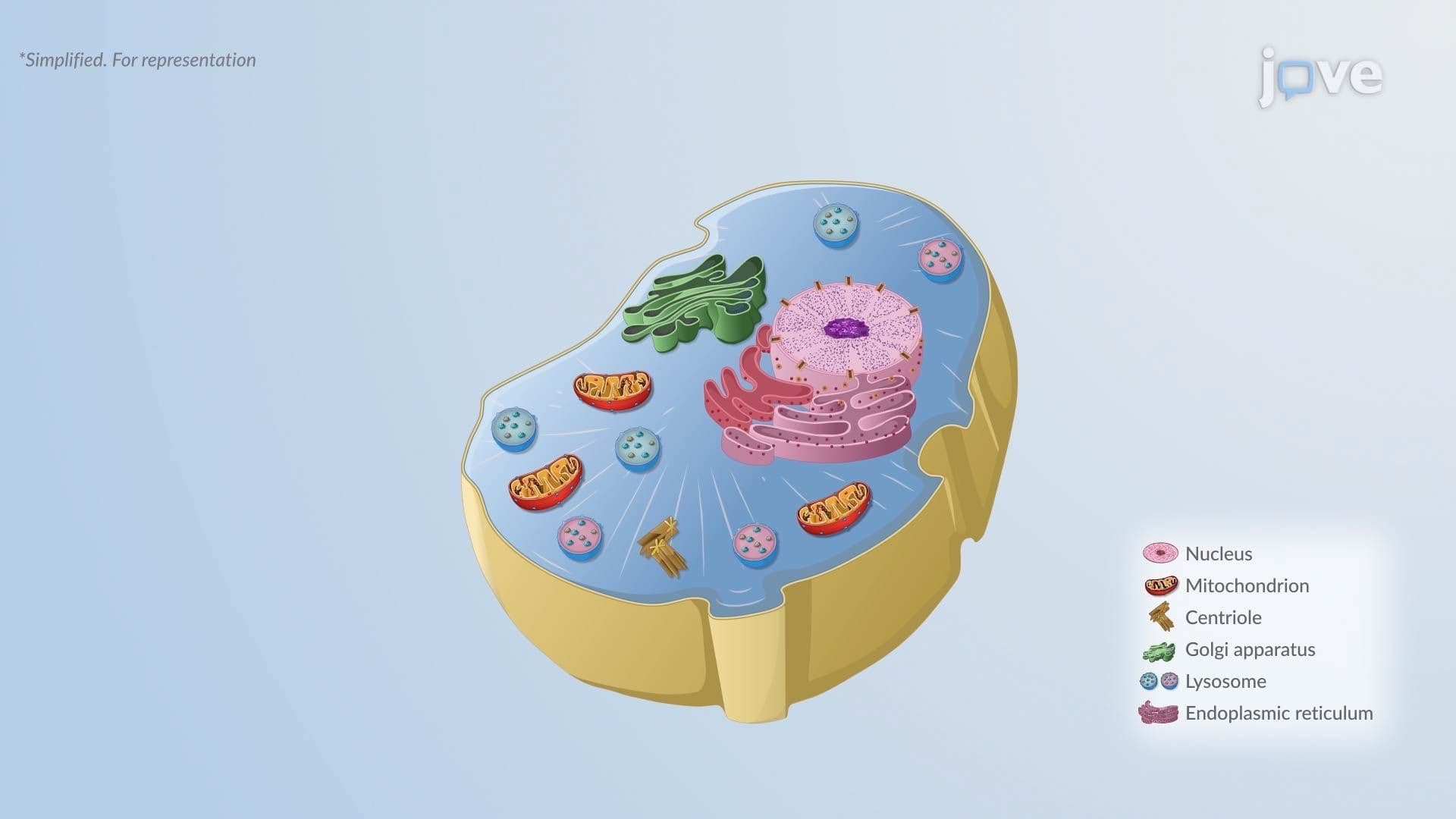
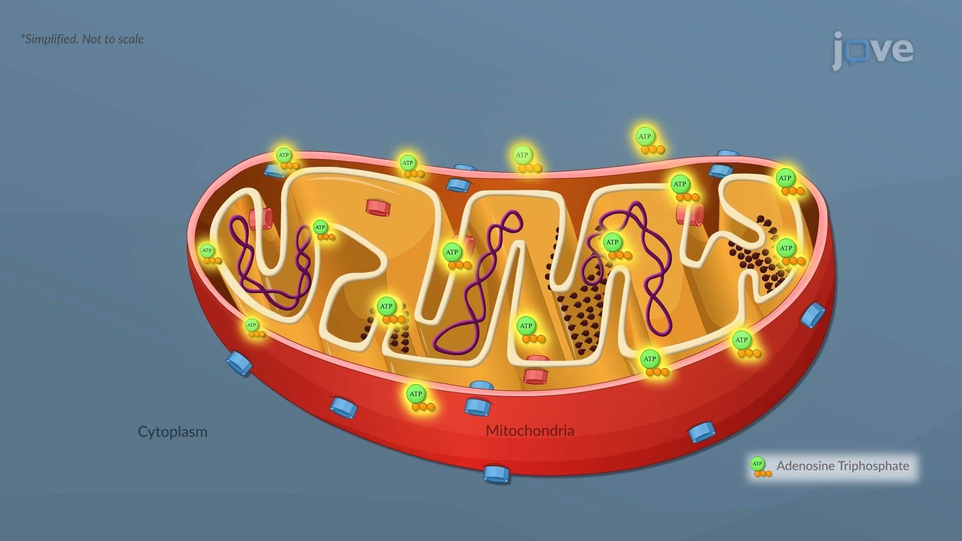
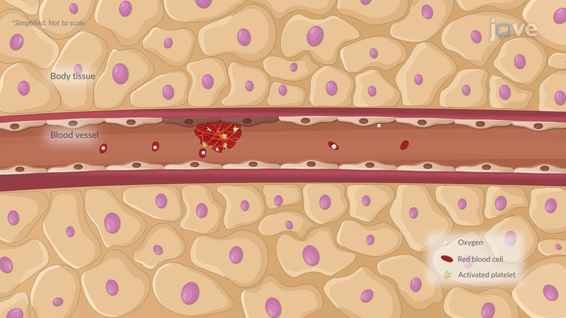
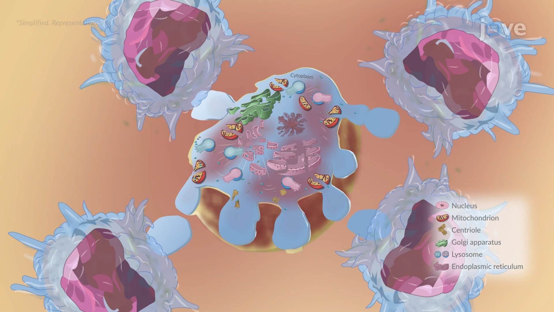
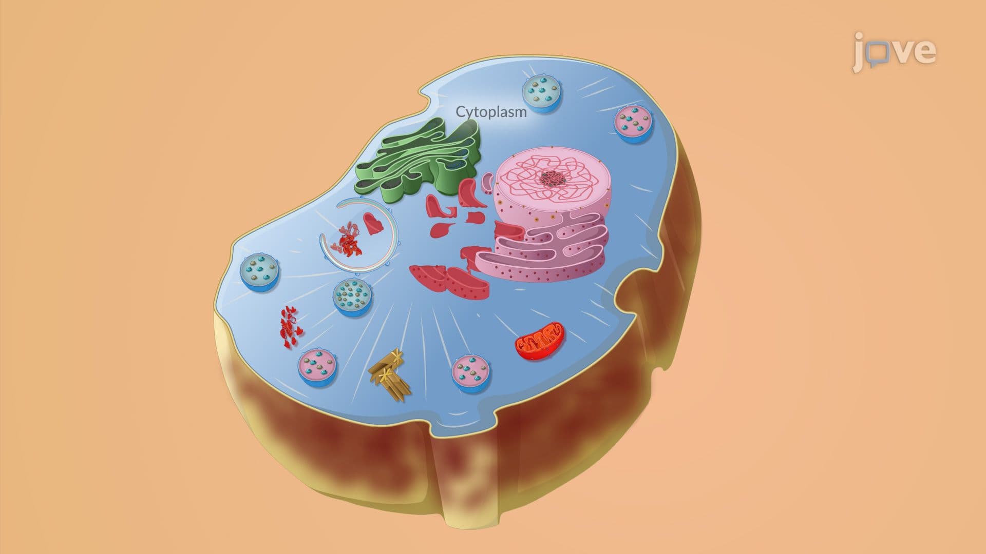
Necrosis follows a predictable sequence. The injury first disrupts cellular energy production, leading to ATP depletion. Without energy, ion pumps fail, and water rushes into the cell, causing it to swell. As the cell membrane loses its integrity, lysosomal enzymes escape into the cytoplasm and begin digesting the cell from the inside out, a process called autolysis.
The nucleus undergoes three distinct, identifiable changes:
These nuclear changes are key identifiers under a microscope and are frequently tested in college-level biology and pathophysiology courses, as well as on the MCAT and USMLE Step 1.
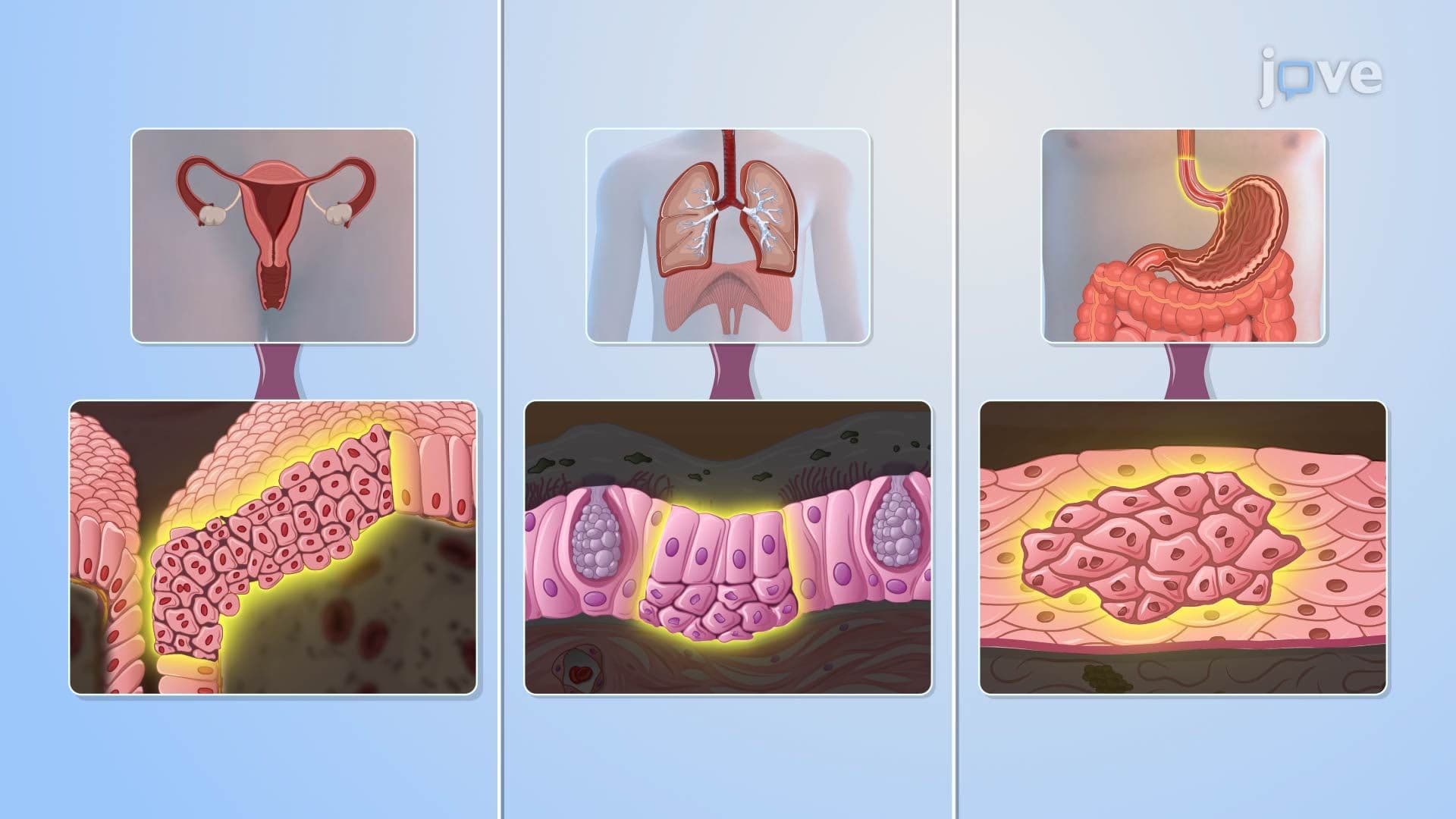
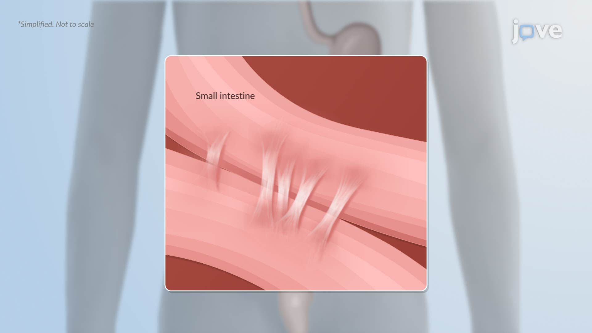
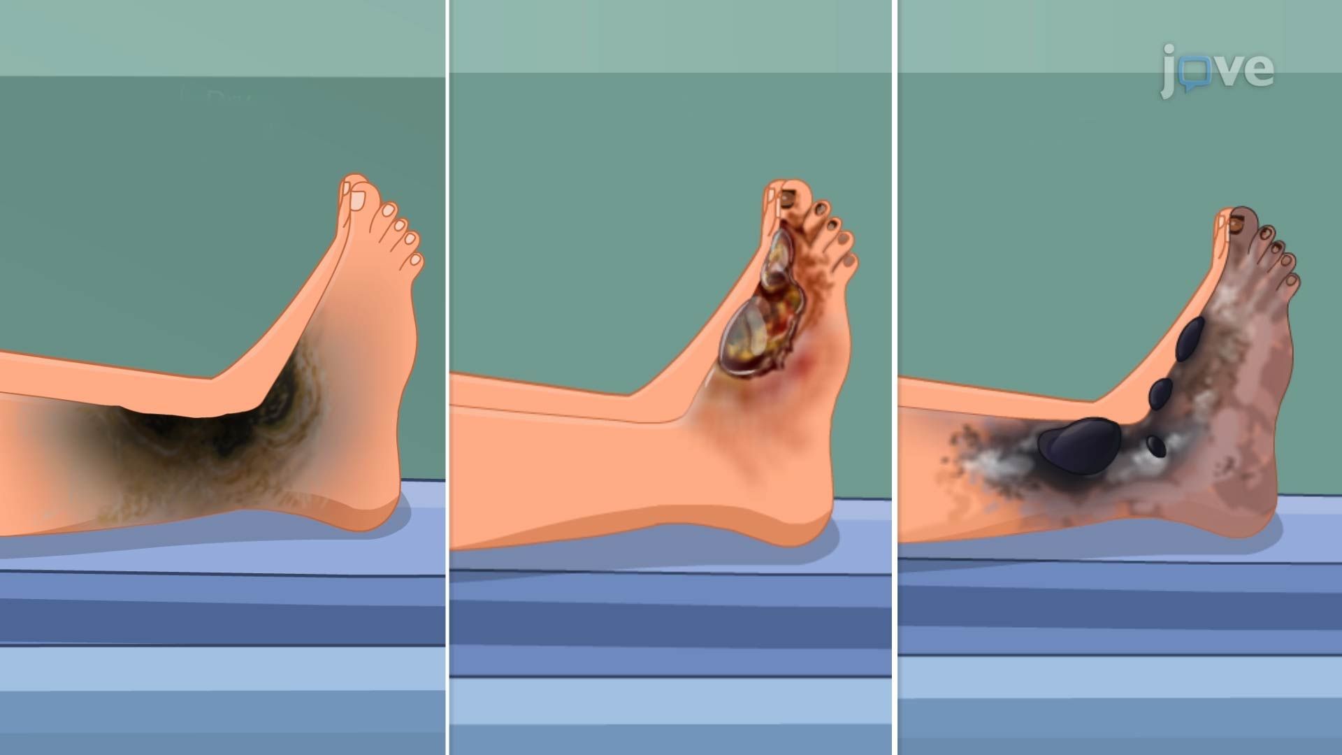
One of the most clinically useful aspects of studying necrosis is recognizing how different types produce distinct tissue changes. This directly supports answering exam questions about what necrotic tissue *looks like*, and why.
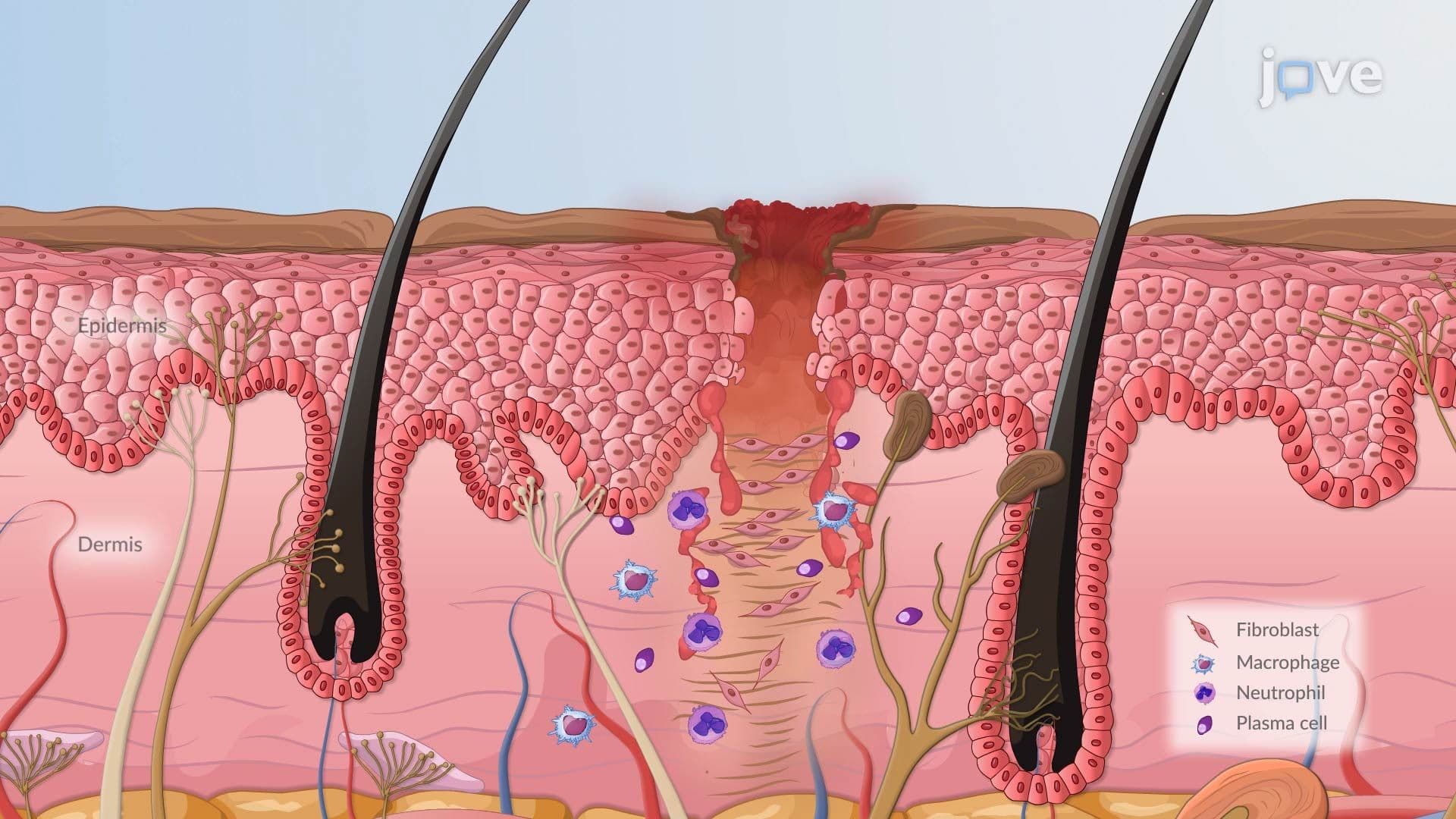
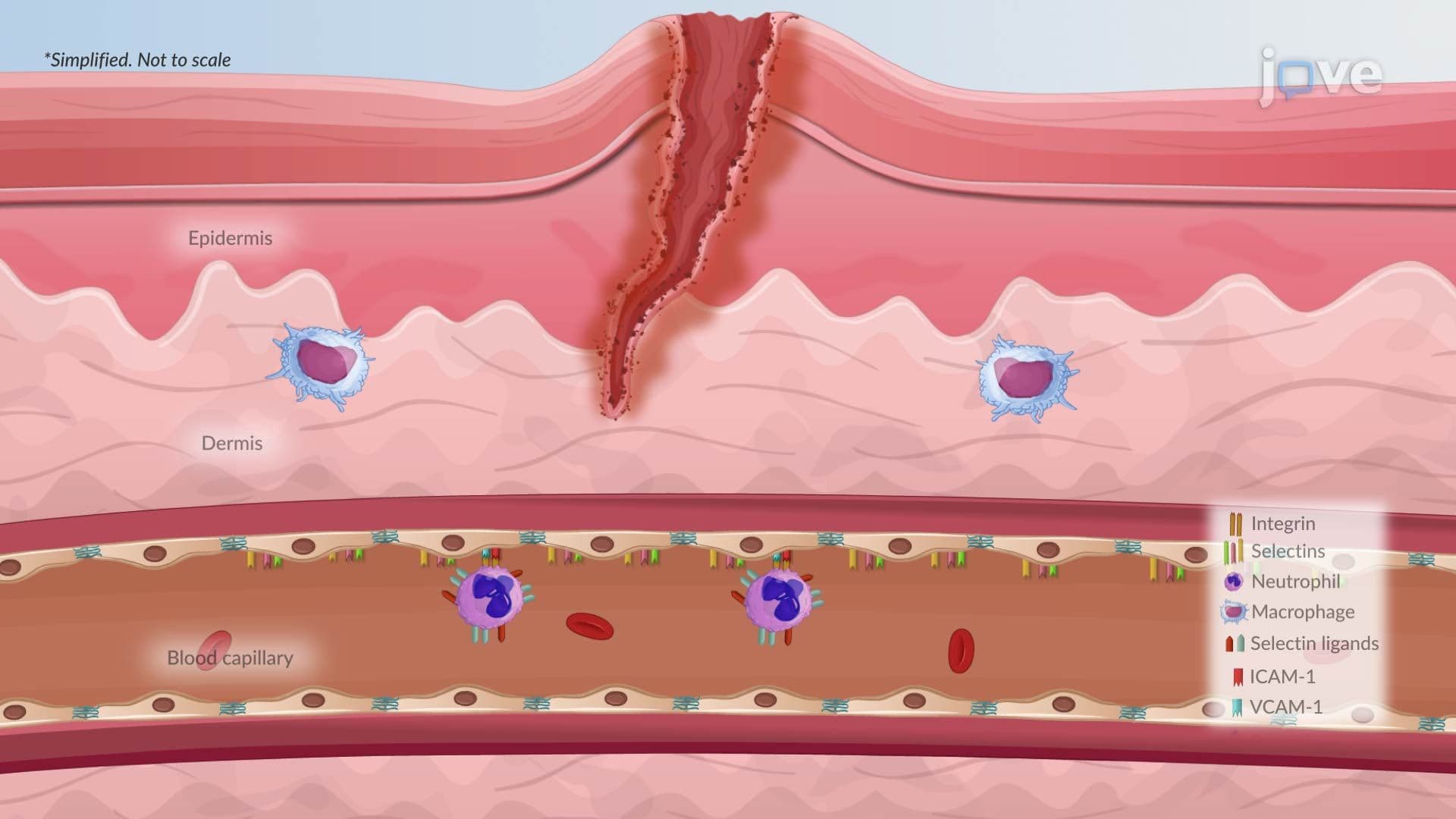
When a cell ruptures during necrosis, it releases damage-associated molecular patterns (DAMPs), internal signals that alert the immune system. Neutrophils and macrophages flood the area to remove debris, initiating the inflammation and repair response. This is a critical distinction from apoptosis, which does *not* trigger inflammation.
In AP Biology and introductory college pathophysiology courses, students are expected to connect necrosis to broader topics like cellular adaptation, hemodynamic disorders, and genetics and disease. Recognizing necrosis as a gateway concept helps students understand how conditions ranging from myocardial infarction to gangrenous diabetic ulcers develop at the tissue level, making it a high-yield topic across multiple US standardized exams and course curricula.
Related Micro-courses