Loading video…

 18 Concepts
 4.3K views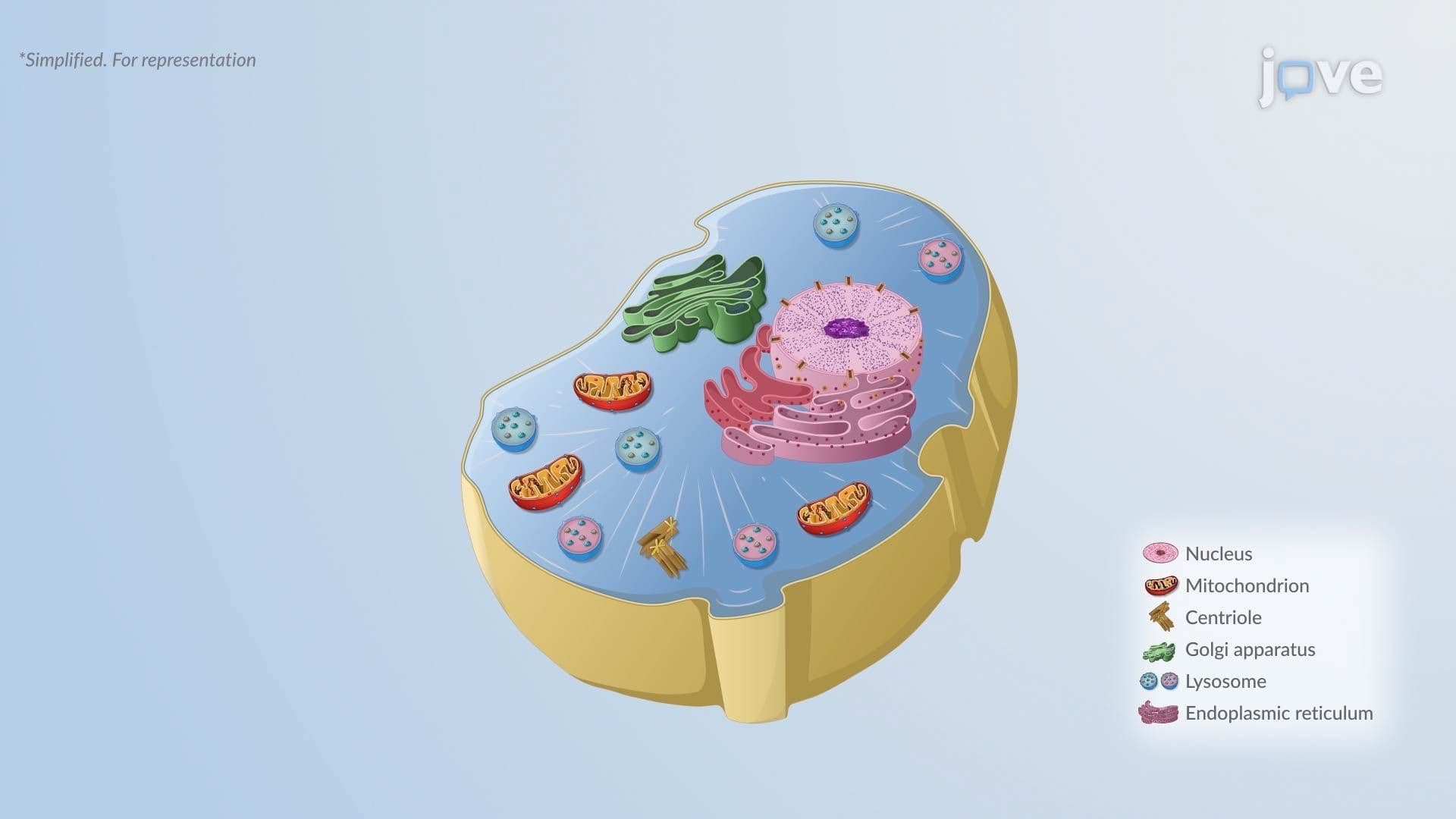

Video Summary: Atrophy in Cellular Adaptation I
Did you know your muscles can literally shrink if you stop using them? Atrophy in Cellular Adaptation I explores exactly this phenomenon, when cells reduce in size or number in response to stress, disease, or disuse. Consider American athletes who experience rapid muscle loss after just weeks of immobilization following injury. Watch the full video on JoVE Coach to master this concept with expert-led visuals and step-by-step explanations.
Cells are not static, they constantly respond to the demands placed on them. Atrophy in Cellular Adaptation I refers to the reduction in cell size, and in some tissues, cell number, as an adaptive response to physiological change or pathological stress. It is one of four major forms of cellular adaptation, alongside hypertrophy, hyperplasia, and metaplasia. Understanding atrophy is foundational for any student studying biology, anatomy, or pre-health sciences, and it appears frequently on exams ranging from AP Biology to the MCAT and USMLE Step 1.
Not all atrophy signals disease. Physiological atrophy occurs naturally and is a normal part of development. The clearest example is thymic involution, the thymus gland, critical for immune development in children, gradually shrinks after puberty. This is entirely normal. Similarly, aging adults experience gradual skeletal muscle atrophy, a process called sarcopenia, which is a major concern in geriatric medicine across the United States.
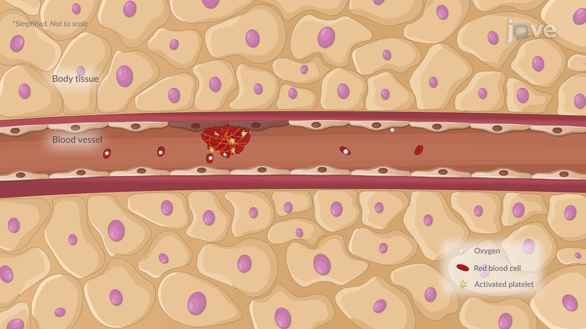
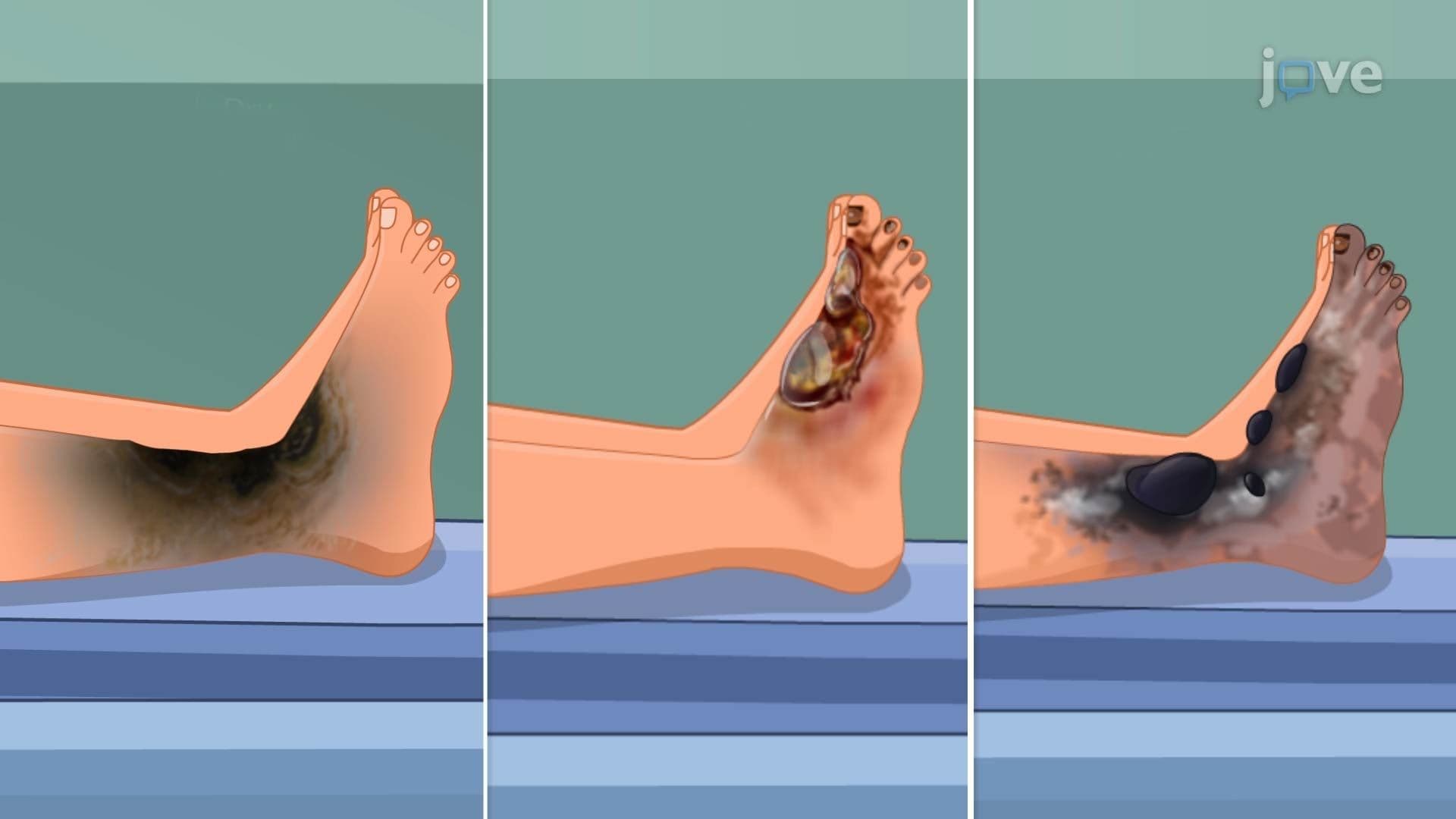
Pathological atrophy, by contrast, is driven by harmful stressors. Causes include chronic malnutrition, nerve damage (denervation atrophy), prolonged bed rest, reduced blood supply (ischemia), and loss of hormonal stimulation. For instance, patients recovering in US hospital ICUs after extended immobility commonly develop disuse atrophy in limb muscles, requiring intensive physical rehabilitation.
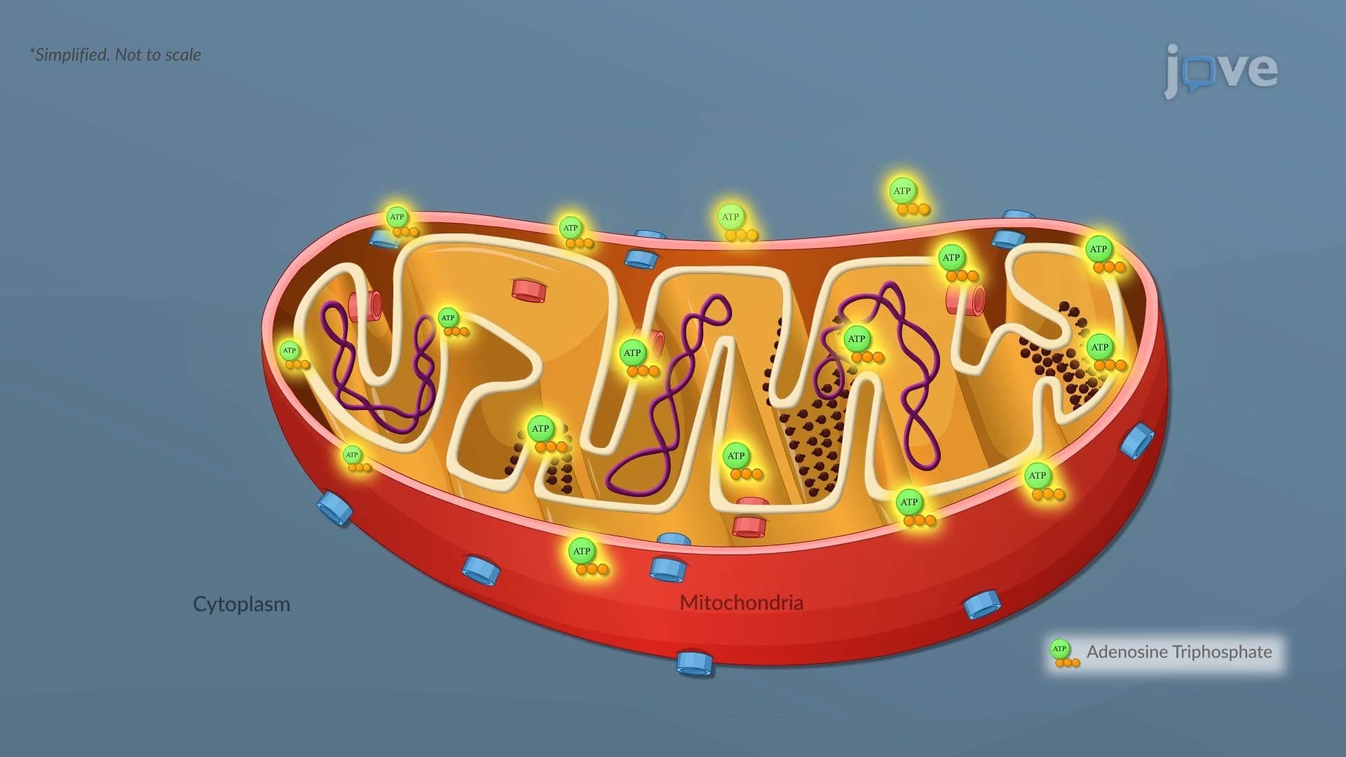
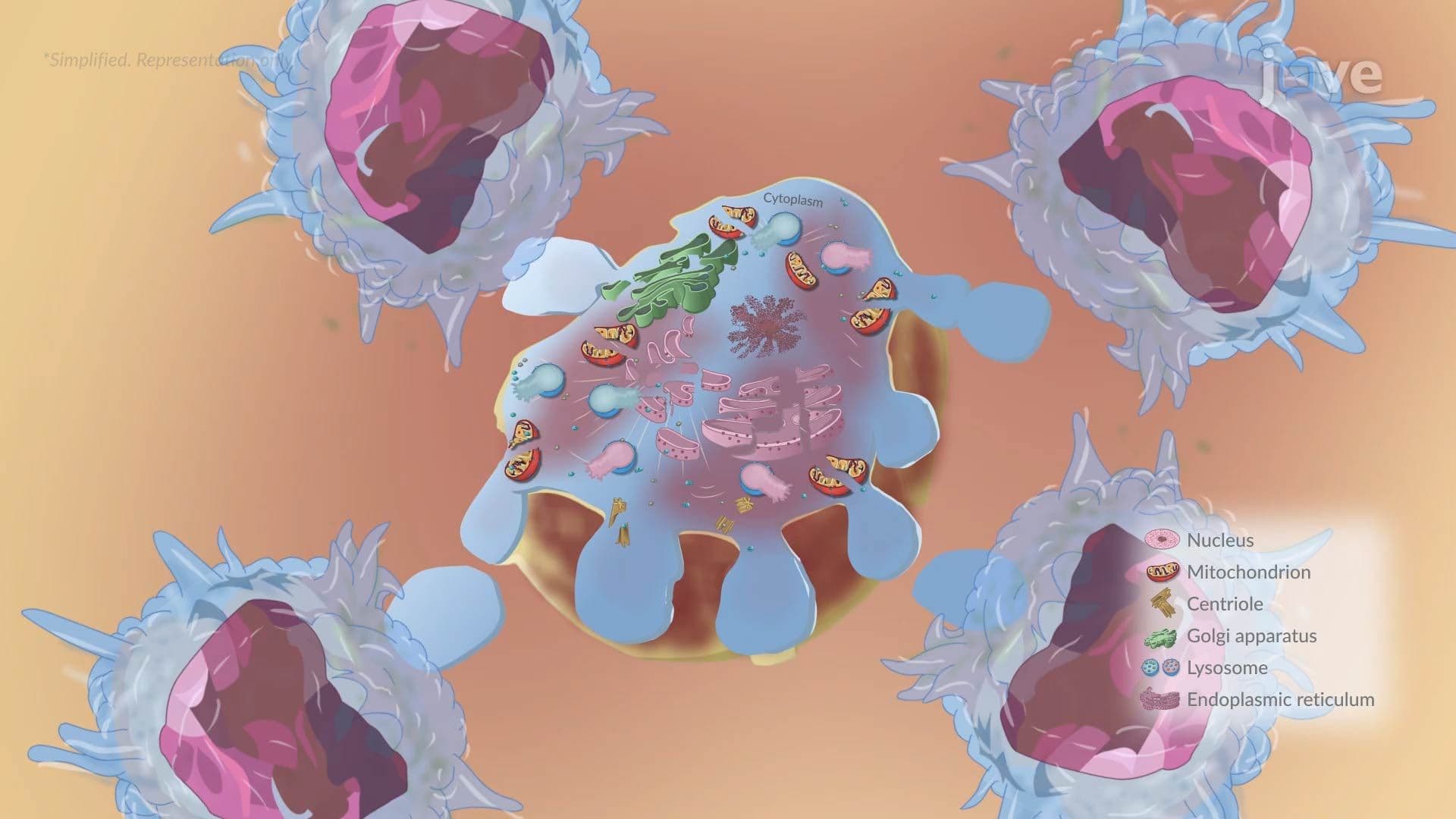
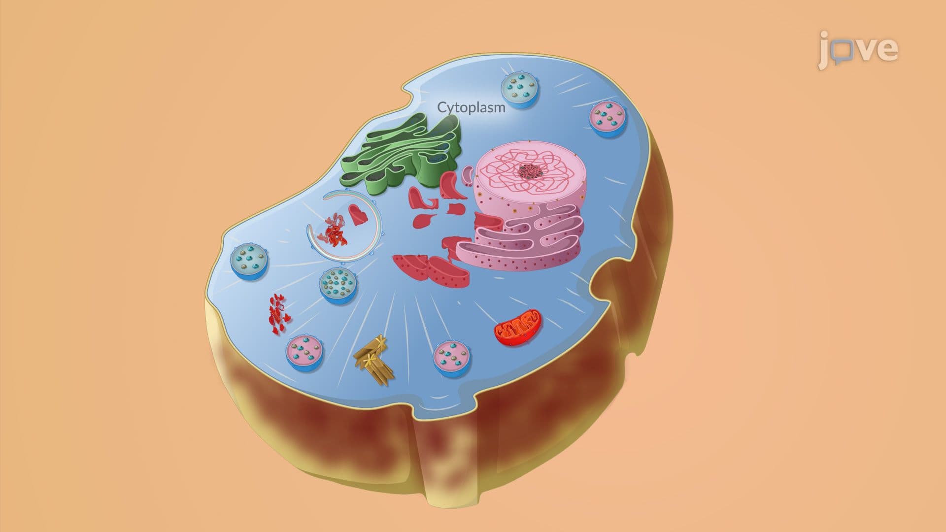
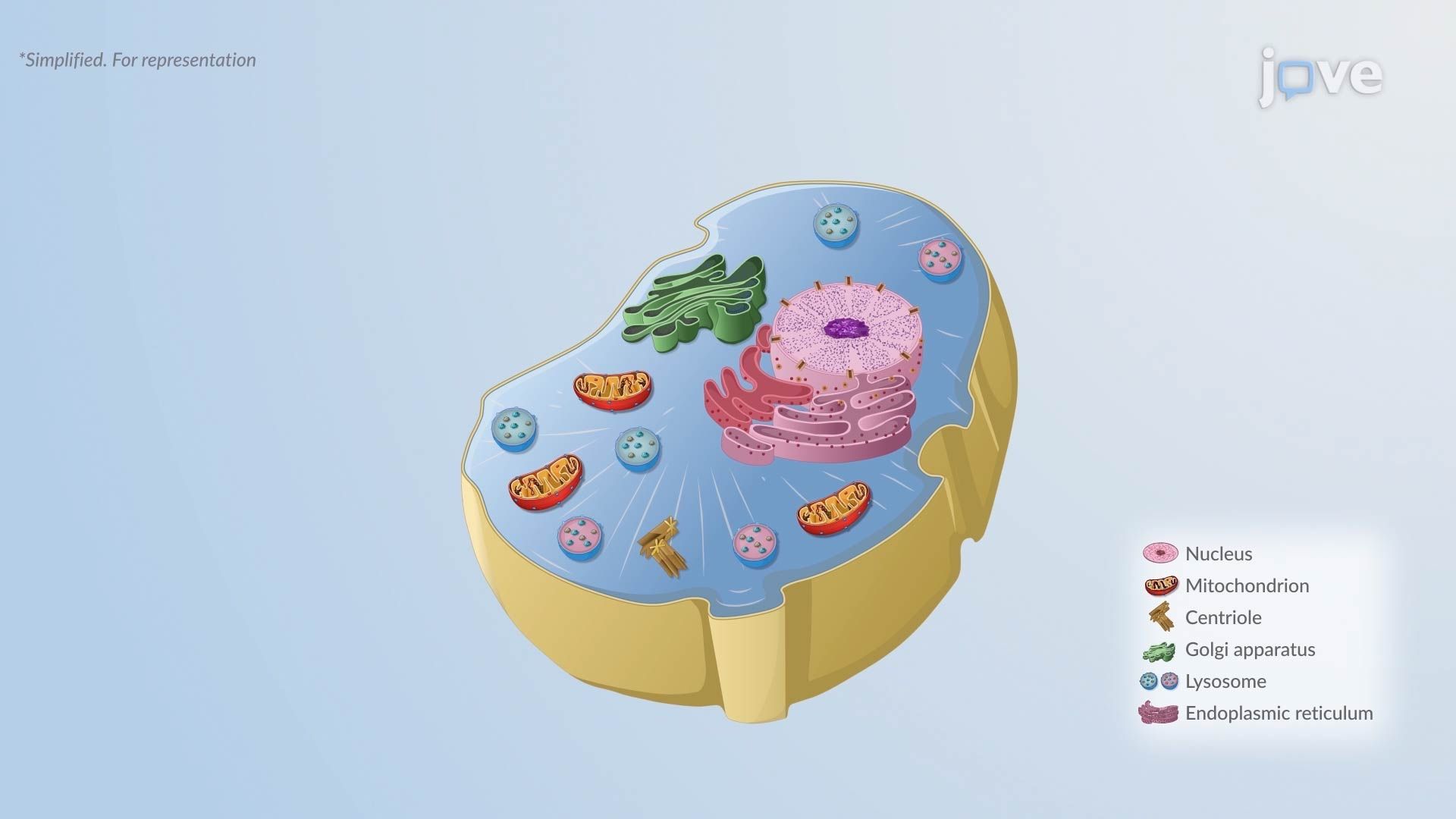
At the cellular level, atrophy is not simply about getting smaller, it reflects a deliberate downregulation of metabolic activity. Atrophic cells contain fewer organelles, particularly mitochondria and rough endoplasmic reticulum (RER). Since mitochondria produce ATP and RER handles protein synthesis, their reduction means the cell consumes less energy and produces fewer proteins. This is the cell's way of surviving on limited resources.
A key process driving atrophy is autophagy, literally "self-eating." During autophagy, the cell packages damaged or unnecessary organelles into structures called autophagosomes, which then fuse with lysosomes for degradation. This recycling process is especially active during starvation or chronic stress. Importantly, some autophagic activity produces lipofuscin, a yellow-brown pigment that accumulates in atrophic cells and is considered a hallmark of cellular aging. Pathologists in the US often note lipofuscin deposits during tissue examination.
Brain atrophy is one of the most clinically significant forms of pathological atrophy studied in US medical and pre-medical curricula. Chronic cerebral ischemia, often resulting from atherosclerosis of the cerebral arteries, deprives neurons of oxygen and nutrients, leading to progressive loss of brain tissue. This is distinct from, but related to, stroke. Conditions such as Alzheimer's disease also produce characteristic patterns of cortical atrophy visible on MRI scans used in American clinical practice.
Understanding how atrophy connects to broader disease mechanisms, including hemodynamic disorders, inflammation and repair, and genetics and disease, gives students a powerful framework for answering systems-level questions on college midterms, MCAT passages, and USMLE-style clinical vignettes. Mastering this concept now builds the vocabulary and reasoning skills needed for advanced pathophysiology courses.
Related Micro-courses