Loading subject page…

4,341 views

Video Summary: What Is Cellular Injury I
Ever wonder why a single blocked artery can destroy heart muscle within minutes? That's cellular injury I basics in action. When cells lose oxygen, a condition called hypoxia, their energy supply collapses, triggering a dangerous chain reaction seen in events like stroke or heart attack across thousands of US hospitals daily. Understanding cellular injury I reveals how disease begins at the microscopic level. Watch the full video on JoVE Coach to master this concept with expert-led visuals and step-by-step explanations.
Every disease, from a common infection to a heart attack, begins with a disruption at the cellular level. Cellular Injury I is the foundational concept in pathophysiology that explains what happens when a cell is pushed beyond its ability to adapt. Cells are remarkably resilient; they constantly adjust to changes in their environment. But when stress is too severe or prolonged, that resilience fails. Understanding *how* and *why* cells get injured is the first step toward understanding how diseases develop, spread, and damage organs.
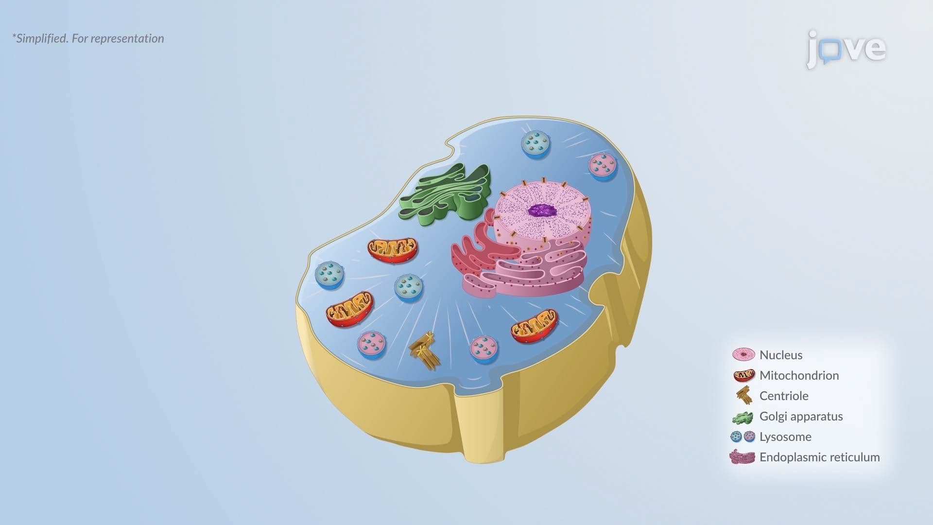
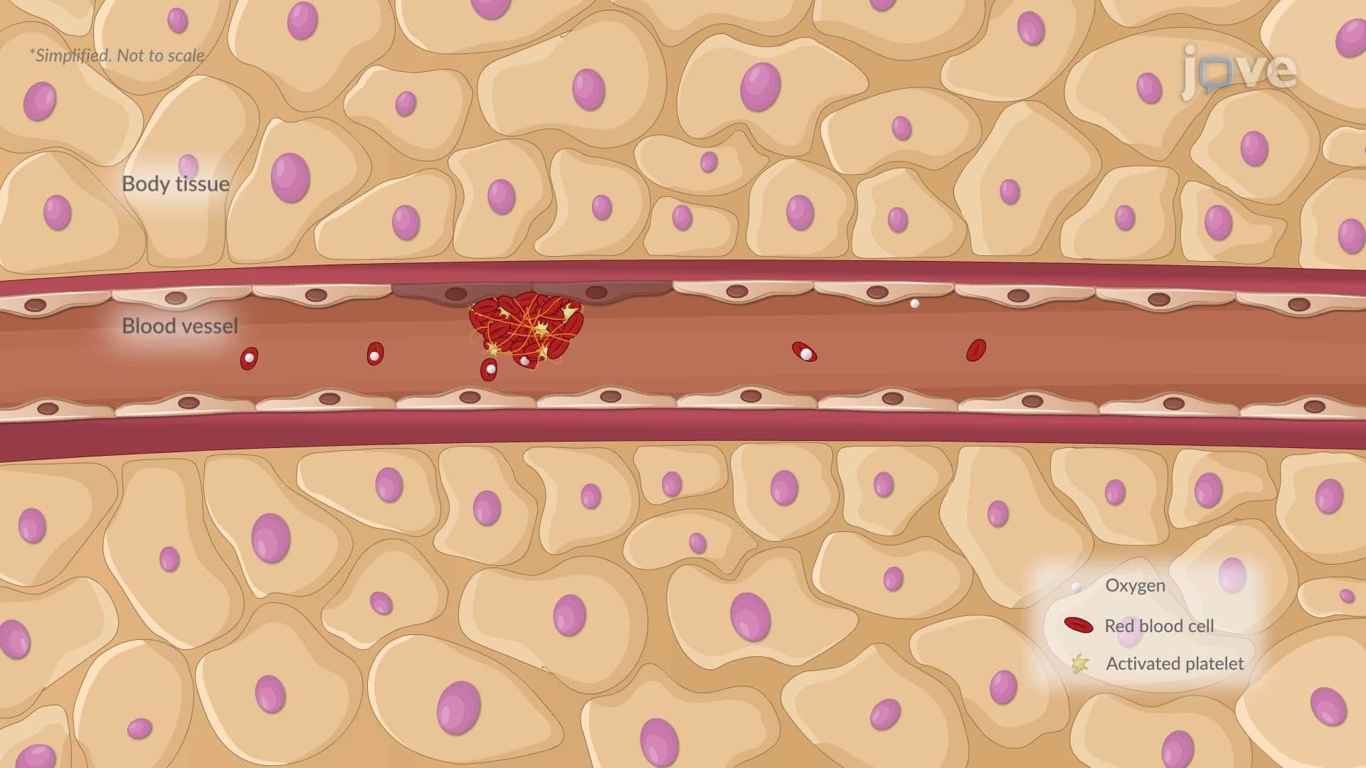
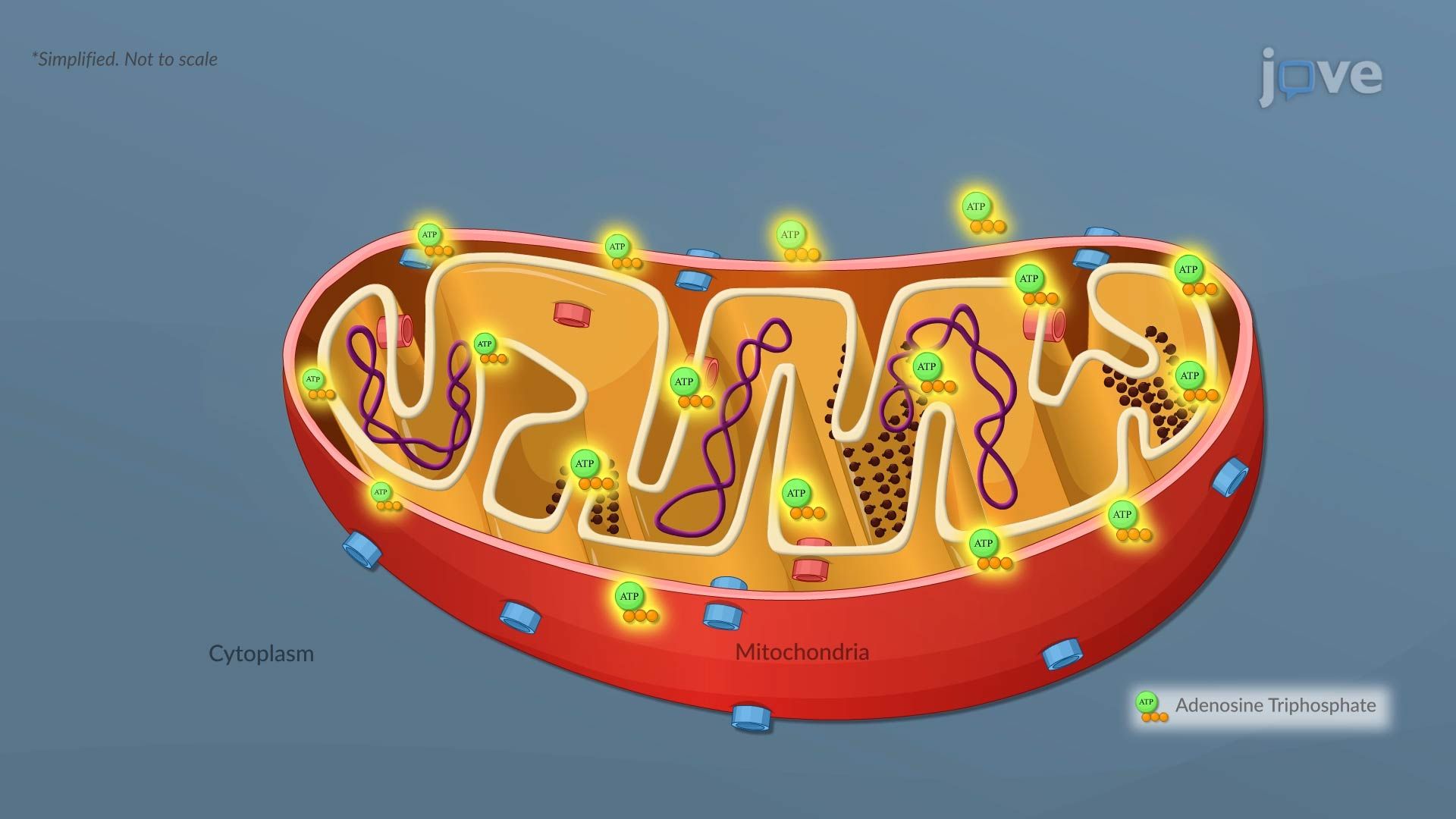
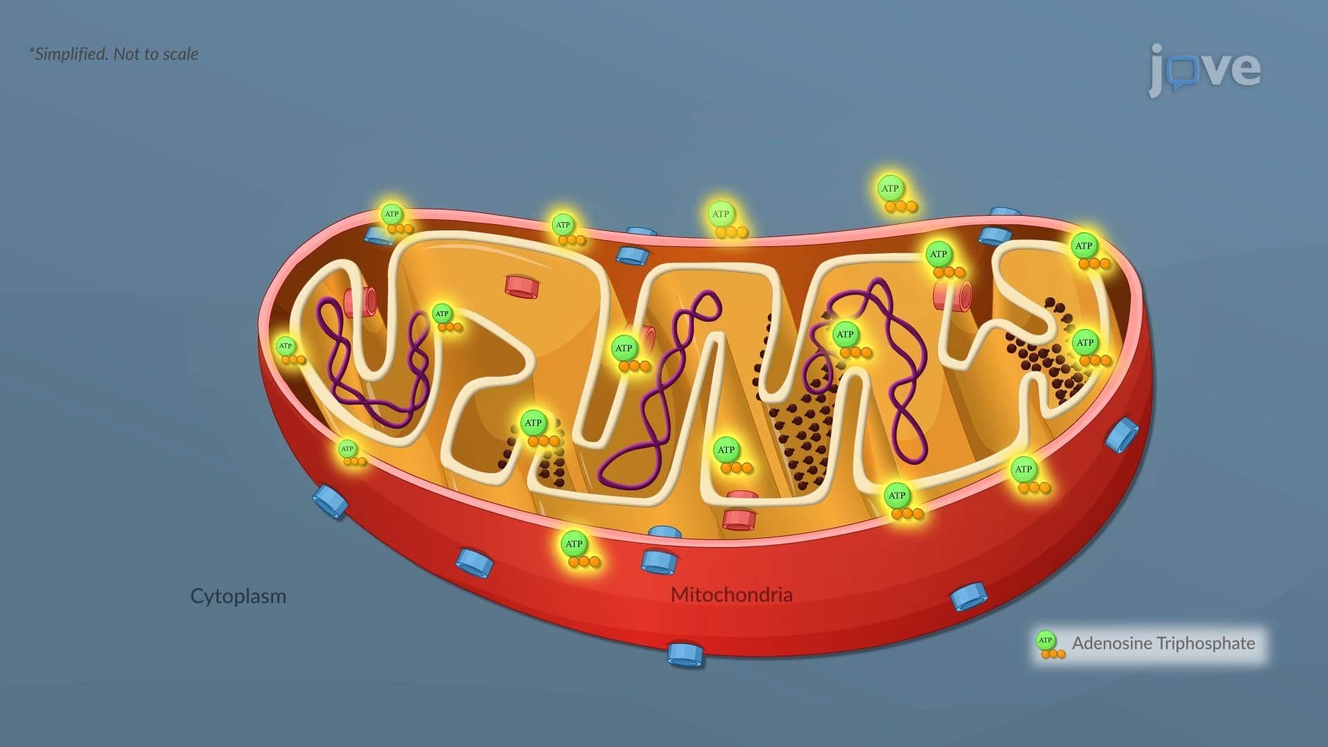
Oxygen is not just necessary for breathing, it is essential for producing ATP, the molecule that powers nearly every cellular function. When oxygen supply drops (hypoxia), mitochondria cannot complete aerobic respiration efficiently, and ATP levels plummet rapidly. This is exactly what happens during a myocardial infarction (heart attack), one of the leading causes of death in the United States. Without adequate ATP, cells lose the energy needed to run their most basic maintenance tasks, setting off a cascade of failures.
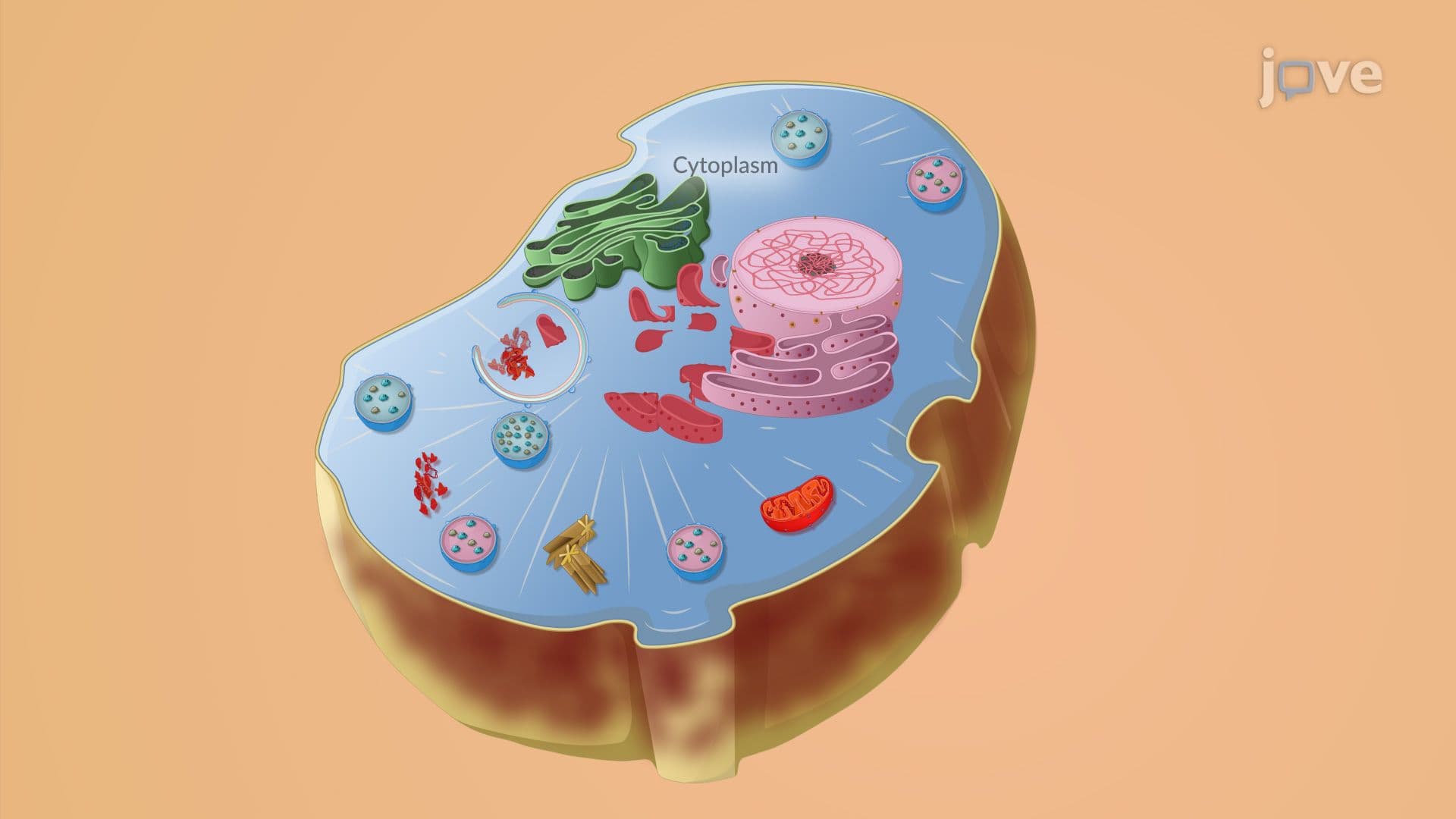
One of the first casualties of ATP depletion is the sodium-potassium ATPase pump, a protein embedded in the cell membrane that normally moves sodium out of the cell and potassium in. Without ATP to fuel it, this pump stops working. Sodium accumulates inside the cell, and because water follows sodium through osmosis, the cell begins to swell, a condition called hydropic change or cellular swelling. At this stage, the injury may still be reversible: restore oxygen and ATP, and the cell can recover. This reversible phase is critical in clinical settings, which is why emergency responders race to restore blood flow in stroke and heart attack patients within narrow time windows.
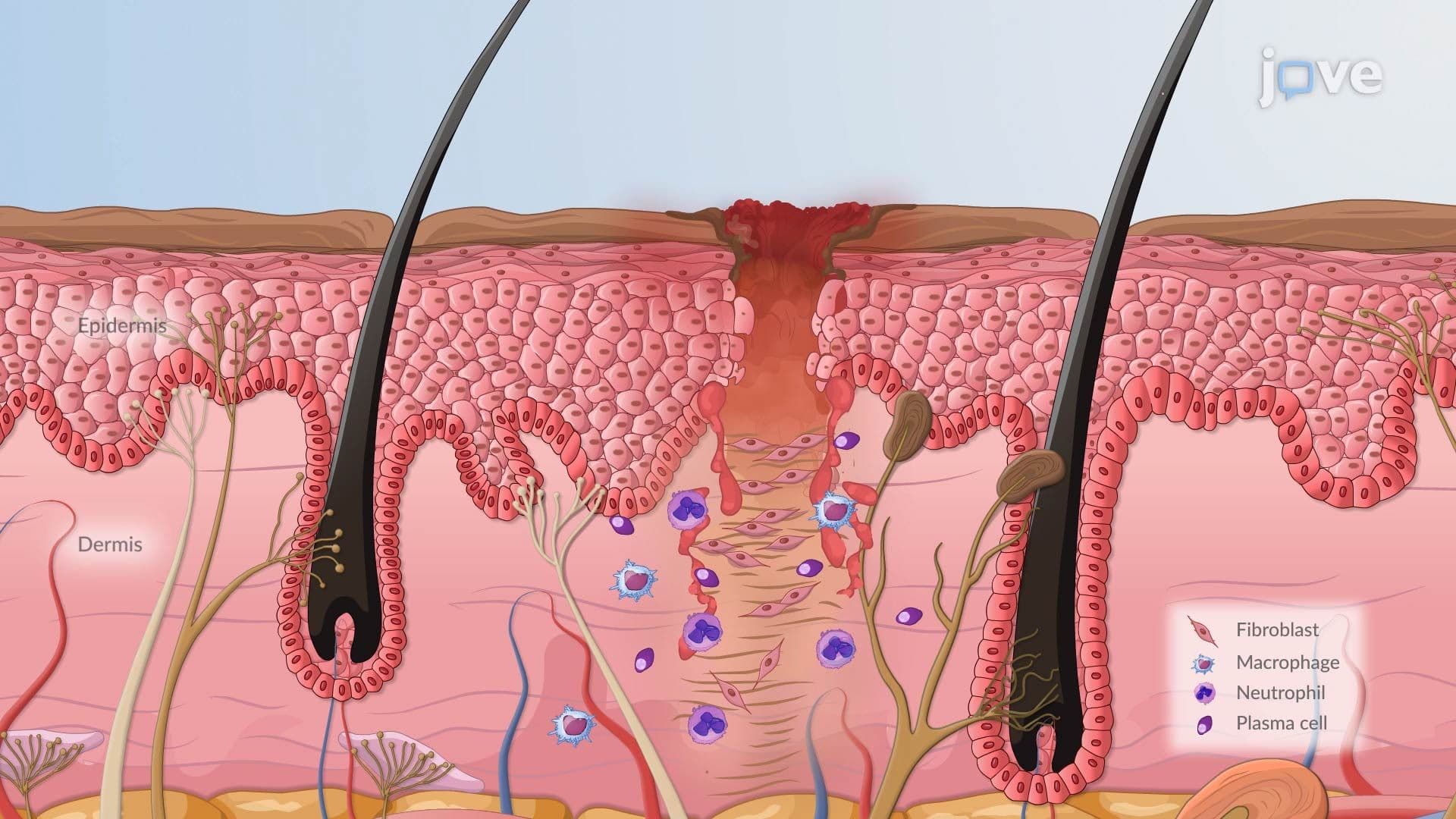
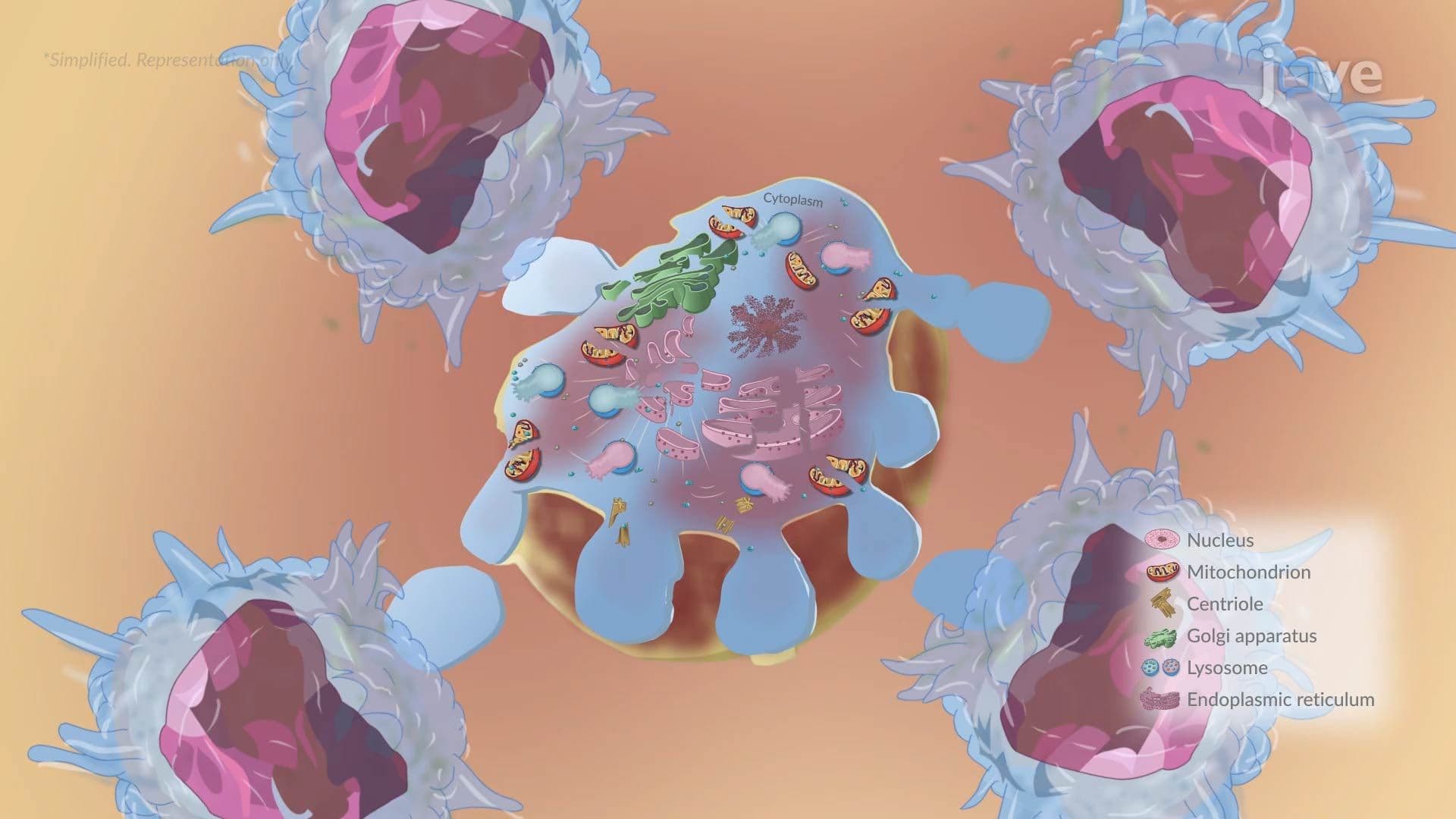
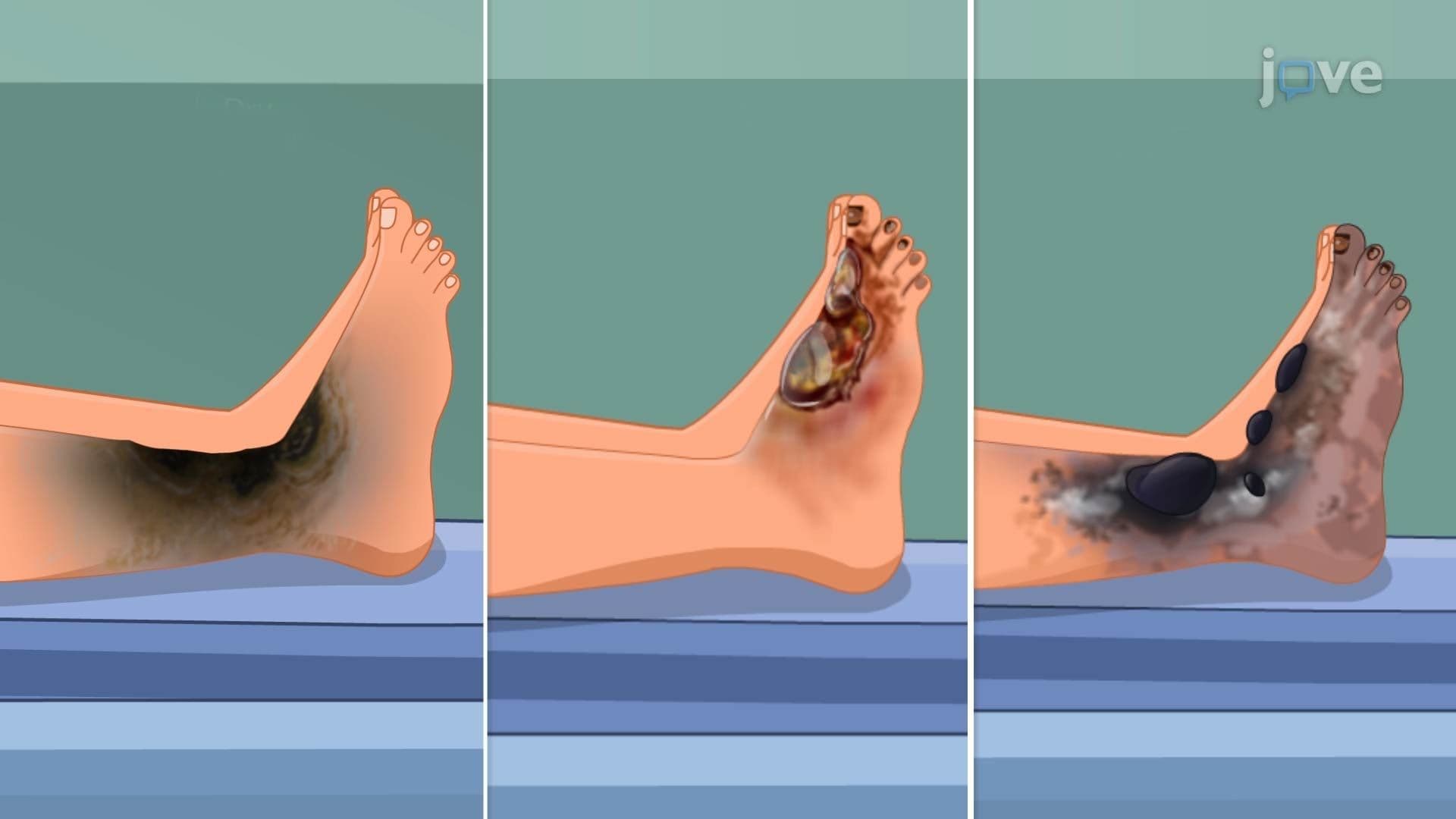
If the stress continues long enough, the cell crosses a threshold into irreversible injury, and cell death becomes unavoidable. Two distinct pathways lead to cell death, and understanding the difference matters both clinically and on exams like the MCAT and USMLE. Necrosis is uncontrolled cell death caused by overwhelming injury, the cell membrane ruptures, cellular contents spill out, and the immune system responds with inflammation. This inflammatory response is what causes the redness, swelling, and pain associated with tissue damage. Apoptosis, by contrast, is a highly organized, programmed process. The cell essentially "self-destructs" in a controlled way, packaging its contents so neighboring cells are not harmed and inflammation is avoided. Apoptosis is normal and necessary; it shapes embryonic development and eliminates pre-cancerous cells. The disruption of apoptosis, when it fails to occur, is actually a hallmark of cancer biology.
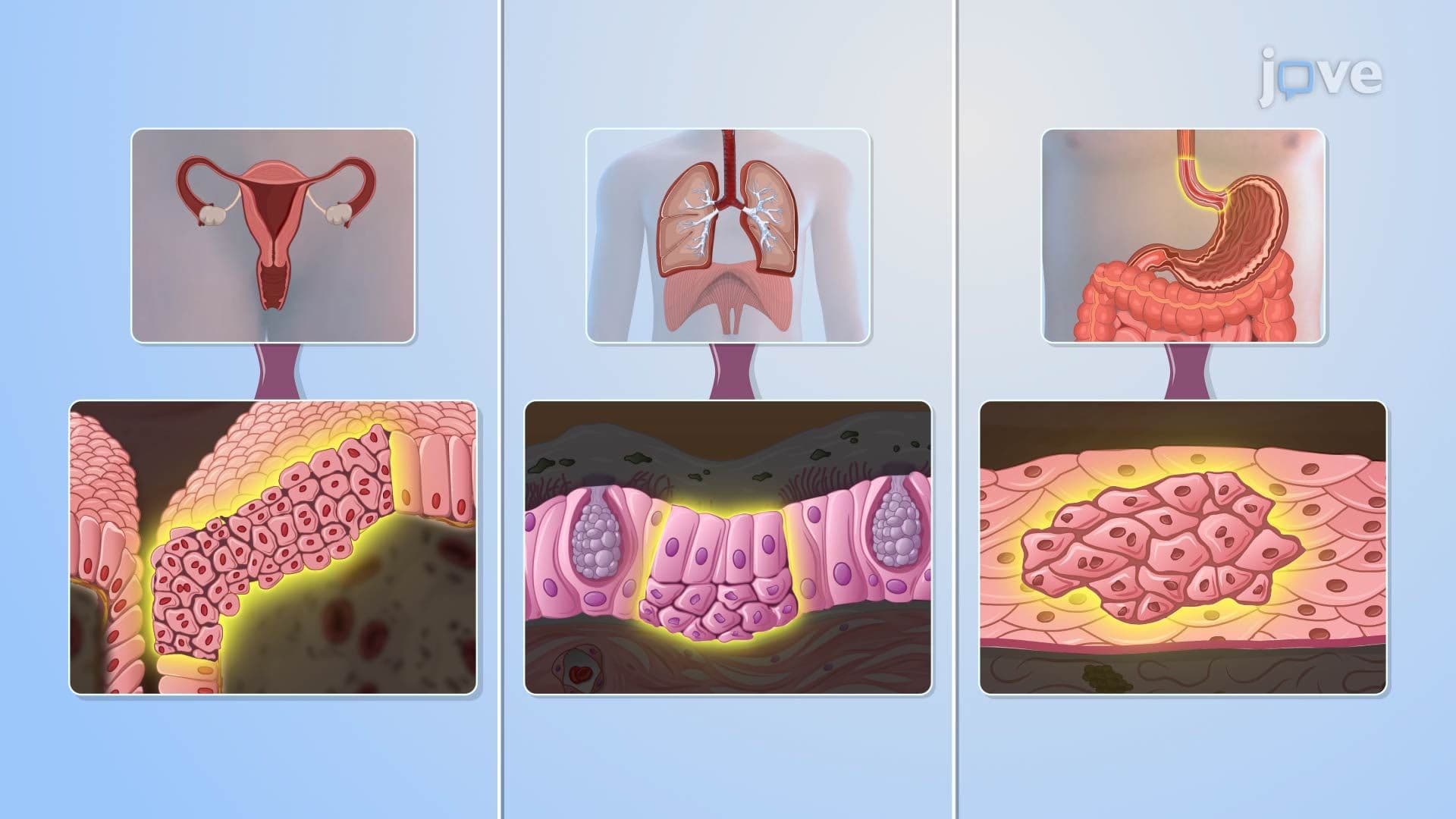
Students in AP Biology, college-level cell biology, anatomy and physiology, and pre-med pathophysiology courses will encounter cellular injury concepts repeatedly. On the MCAT, questions about ischemia, cellular adaptation, and necrosis vs. apoptosis are common. Connecting the molecular (ATP depletion, ion pump failure) to the tissue level (swelling, inflammation, cell death) is exactly the kind of multi-scale thinking these exams reward. Mastering Cellular Injury I gives students a powerful framework for understanding not just disease mechanics, but also how the body attempts to heal and adapt, topics covered in hemodynamic disorders, inflammation and repair, and genetics and disease units.
Related Micro-courses