Loading video…

 11 Concepts
 7K views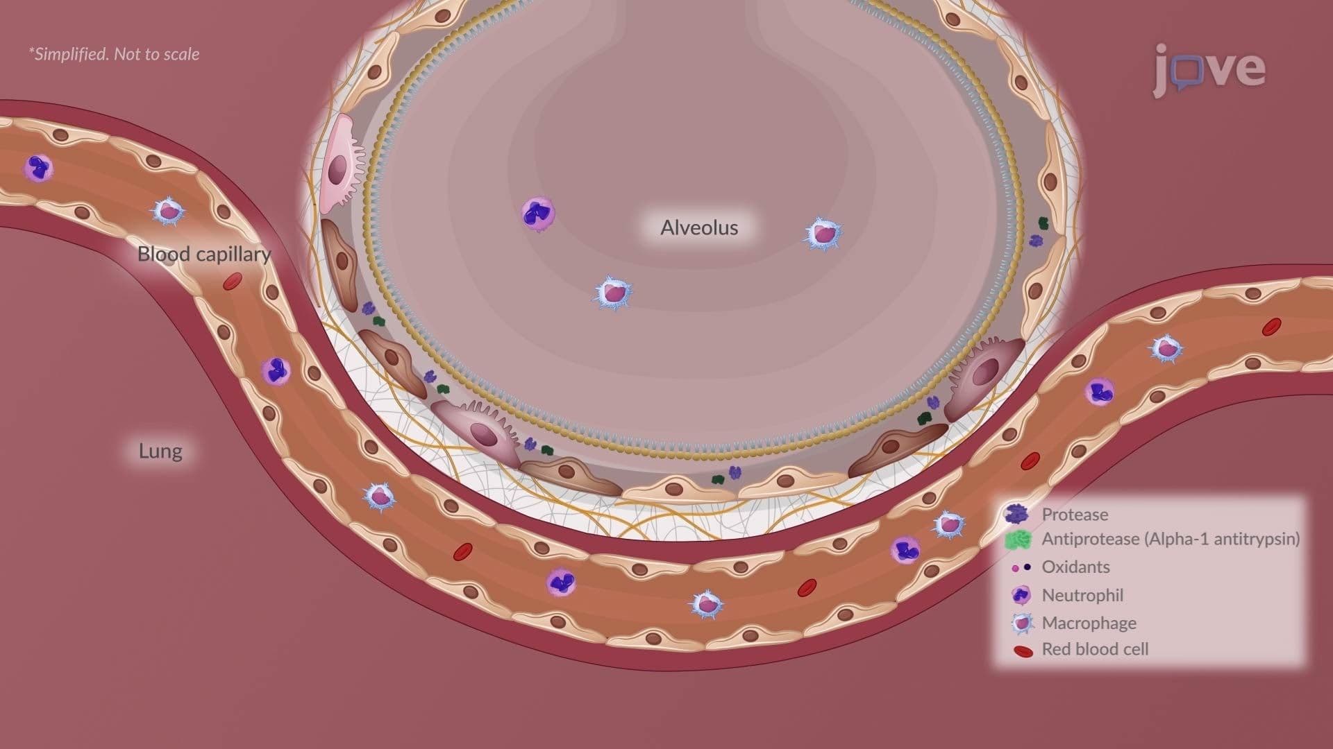

Video Summary: Emphysema in Chronic Obstructive Pulmonary Disease Ii
Did you know that smoking a single pack of cigarettes daily for decades can permanently destroy millions of tiny air sacs in your lungs? Emphysema in Chronic Obstructive Pulmonary Disease II explains exactly how this irreversible damage unfolds, from toxic gas exposure to the collapse of alveolar walls. In the US, over 3 million adults currently live with diagnosed emphysema. Watch the full video on JoVE Coach to master this concept with expert-led visuals and step-by-step explanations.
Emphysema is far more than a breathing problem, it is a slow, irreversible architectural collapse of the lung. As a major phenotype of COPD, emphysema involves the permanent enlargement of distal airspaces caused by the progressive destruction of alveolar walls. Understanding this condition at a mechanistic level is essential for students in AP Biology, college-level anatomy and physiology, and health science programs across the US.
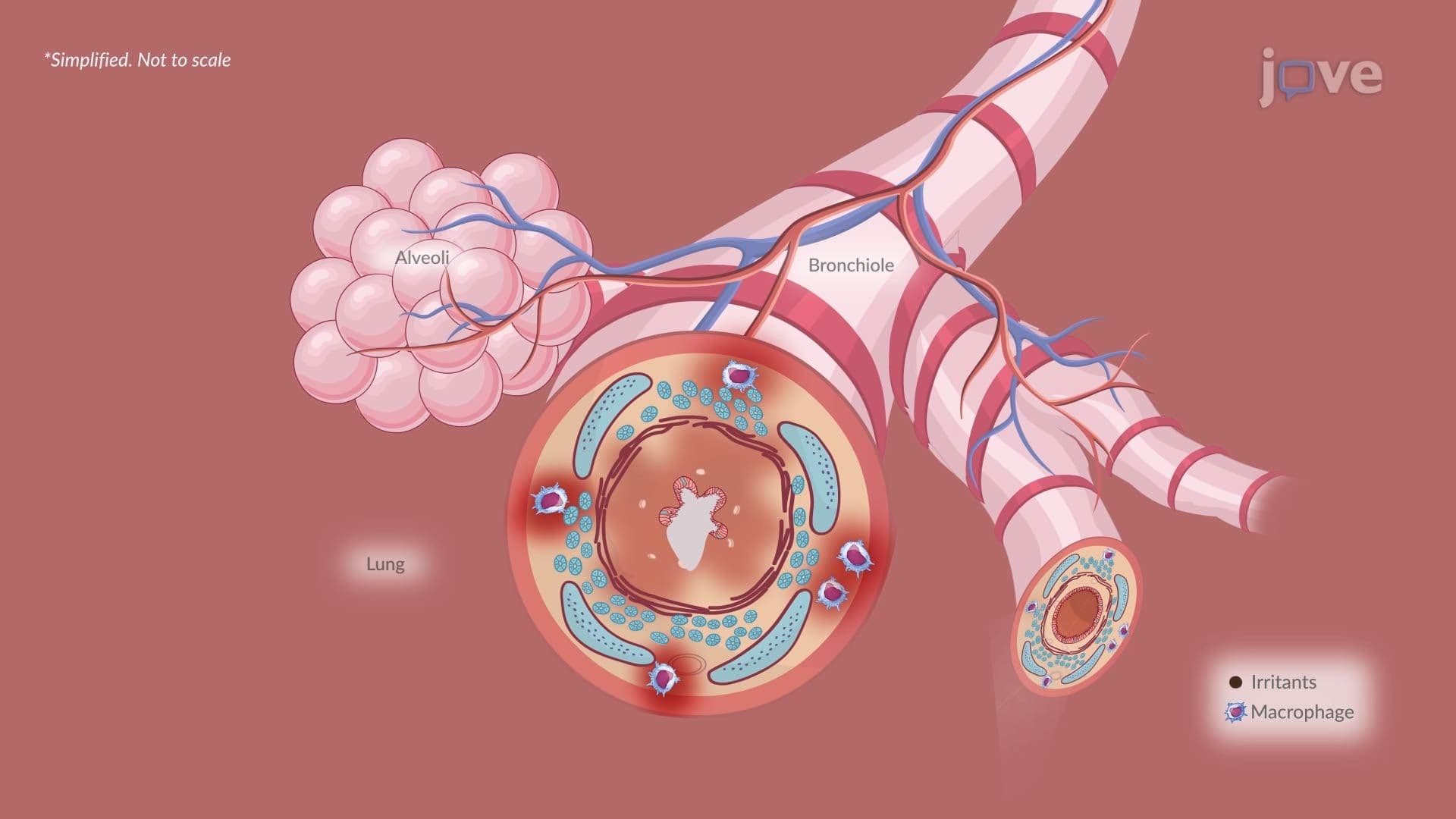
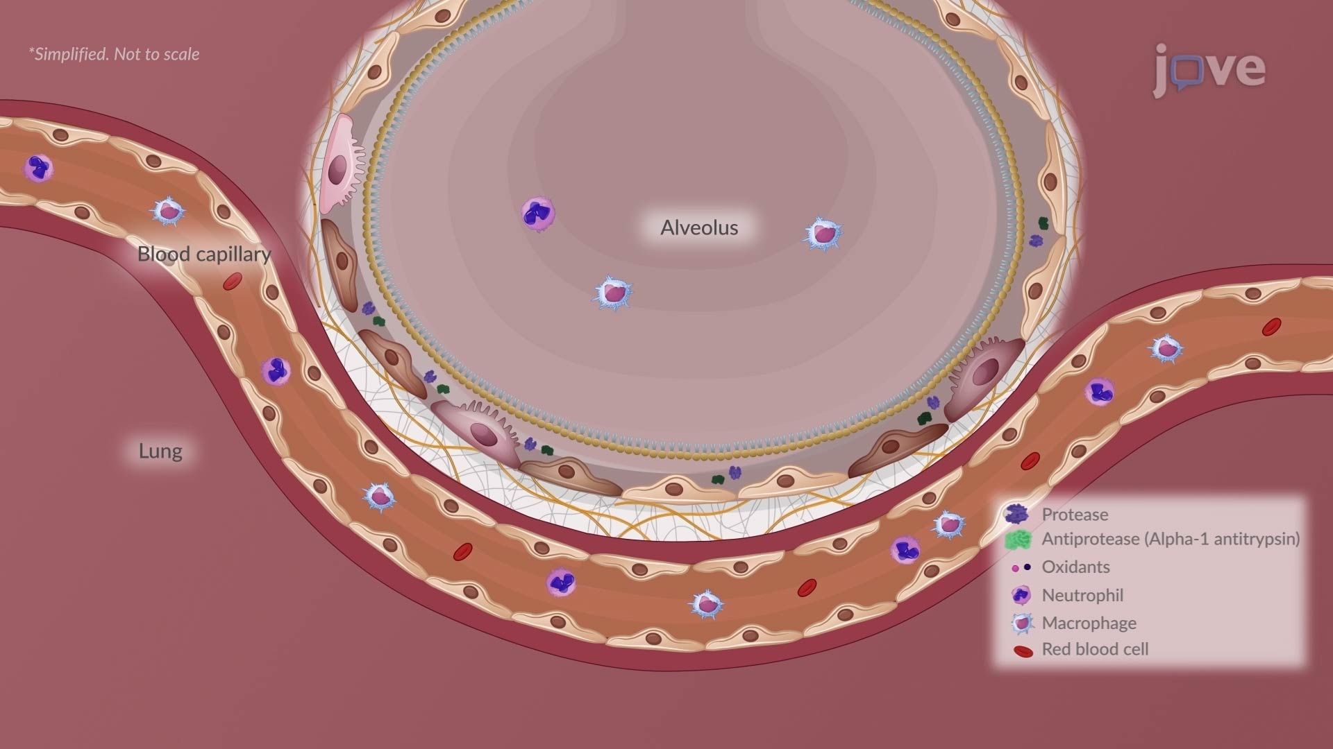
When a person inhales cigarette smoke or other toxic gases over years, the lungs are flooded with harmful oxidants. These oxidants attack a critical defense mechanism: the protease-antiprotease balance. Under normal conditions, α₁-antitrypsin (AAT) neutralizes destructive enzymes like neutrophil elastase, keeping them in check. However, oxidants inactivate AAT, removing this protective brake. Neutrophil elastase then degrades elastin, the protein that gives alveolar walls their stretchiness, along with surrounding connective tissue. The result is the gradual disintegration of the alveolar septa. This cascade is particularly important in students with genetic AAT deficiency, a condition diagnosed in approximately 100,000 Americans, where the protease-antiprotease imbalance occurs even without heavy smoking.
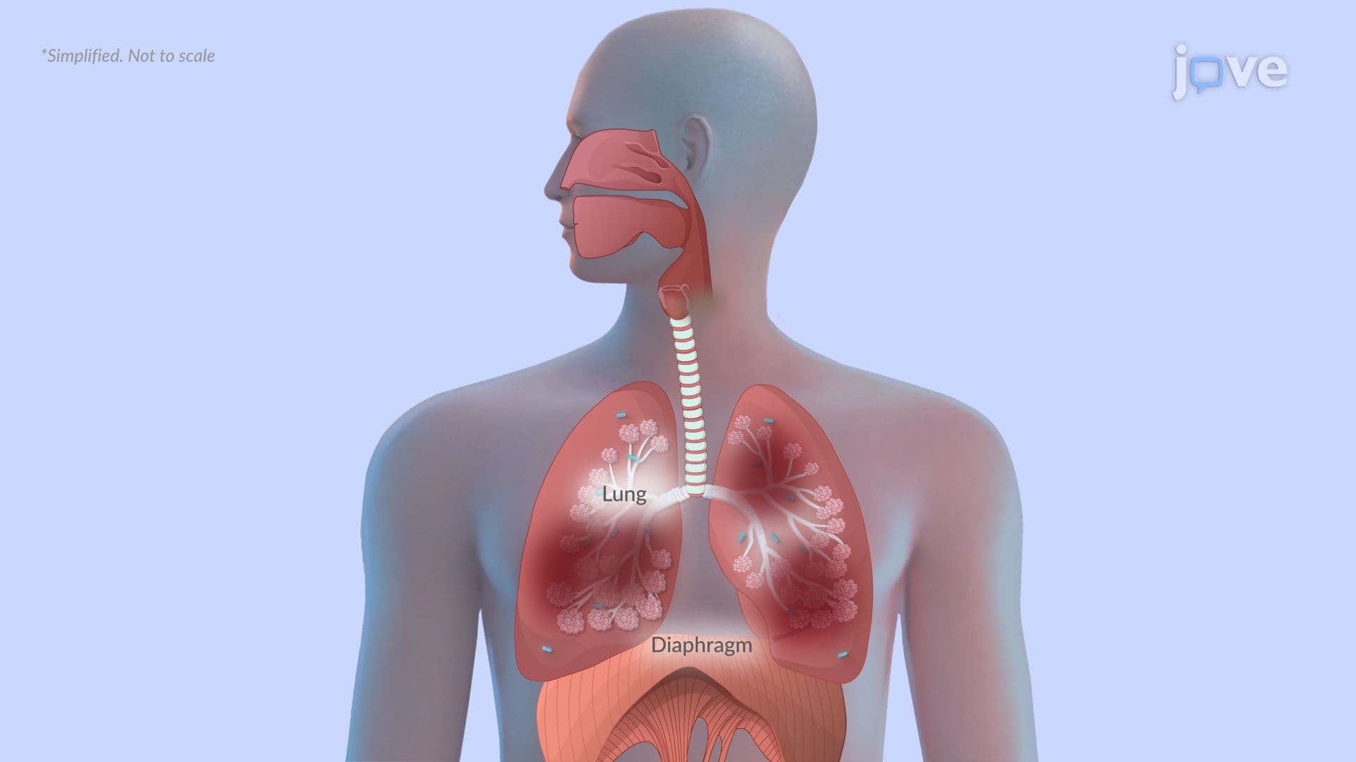
As alveolar walls are destroyed, neighboring air sacs lose their boundaries and merge into large, nonfunctional cavities. When these cavities form deep within the lung parenchyma, they are called bullae. When they form closer to the pleural surface, they are called blebs. Both dramatically reduce the functional surface area available for gas exchange. This is why emphysema patients experience significant ventilation-perfusion (V/Q) mismatch, areas of the lung receive airflow but lack adequate blood flow for oxygen transfer, leading to hypoxemia. In severe US clinical cases, bullae can occupy an entire lung lobe, sometimes requiring surgical intervention called bullectomy.
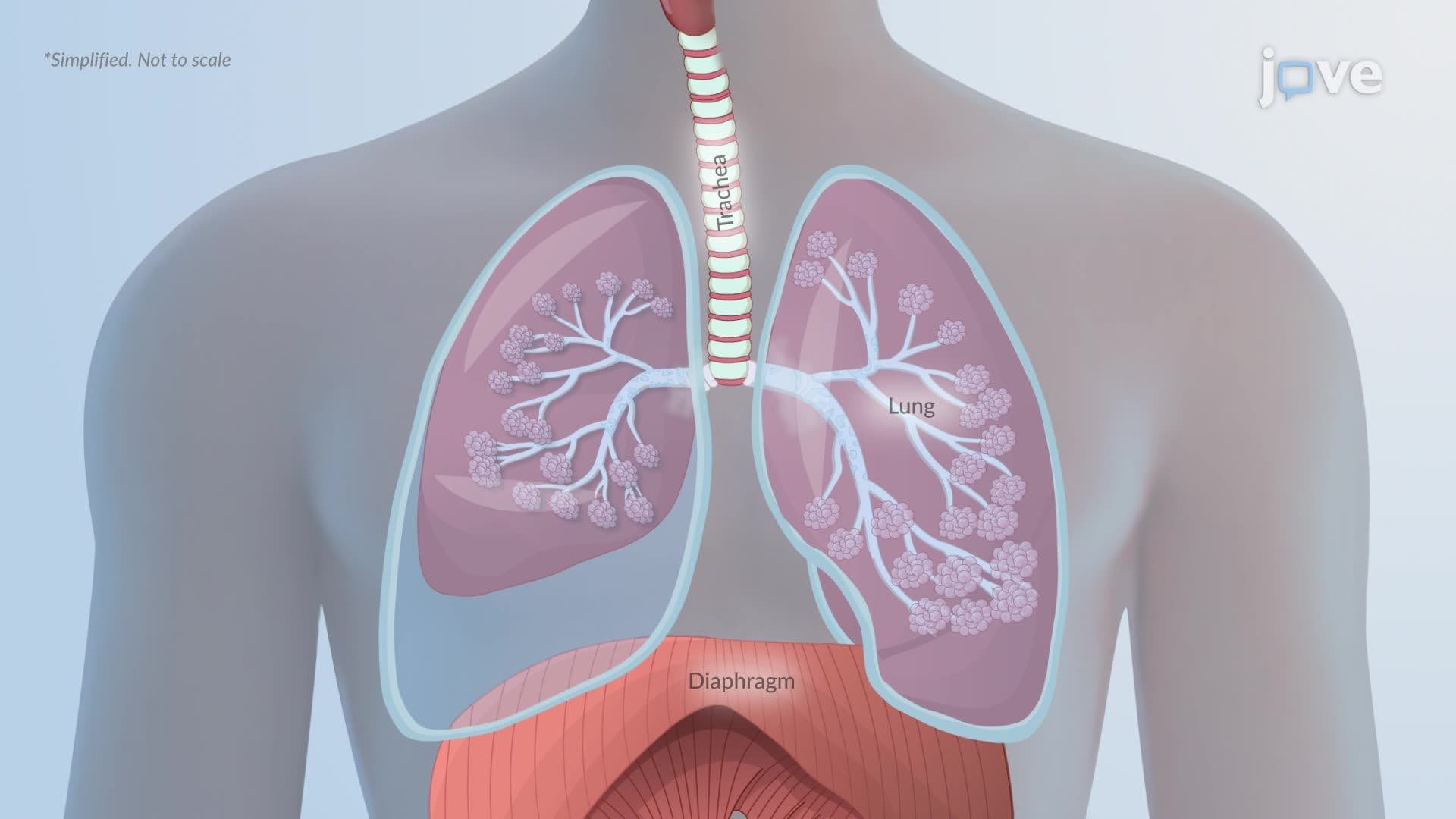
Healthy lung tissue behaves like a stretched rubber band, it naturally recoils during exhalation, pushing air out efficiently. In emphysema, the destruction of elastin fibers eliminates this recoil. Without it, the airways collapse during expiration before air can fully exit, trapping stale, oxygen-depleted air inside the lungs. Over time, this air trapping causes lung hyperinflation, where the lungs remain in a chronically over-expanded state. The diaphragm, which normally domes upward to assist with breathing mechanics, flattens under the pressure of hyperinflated lungs. This compromised position makes every breath require significantly more muscular effort, a phenomenon well recognized in pulmonary rehabilitation programs across US hospitals.
Emphysema pathophysiology is a high-yield topic on the MCAT, USMLE Step 1, NCLEX-RN, and college anatomy and physiology midterms. Questions frequently test students on the mechanism of α₁-antitrypsin deficiency, the physiological basis of air trapping, and how emphysema differs from conditions like asthma, pneumonia, pulmonary embolism, and restrictive lung diseases. Understanding what causes respiratory system disorders at this mechanistic level, rather than simply memorizing symptoms, gives students a significant advantage. It also provides a strong conceptual foundation for exploring related topics such as lung cancer, pulmonary embolism, and how asthma is managed differently from COPD.
Related Micro-courses