Loading video…

 11 Concepts
 7K views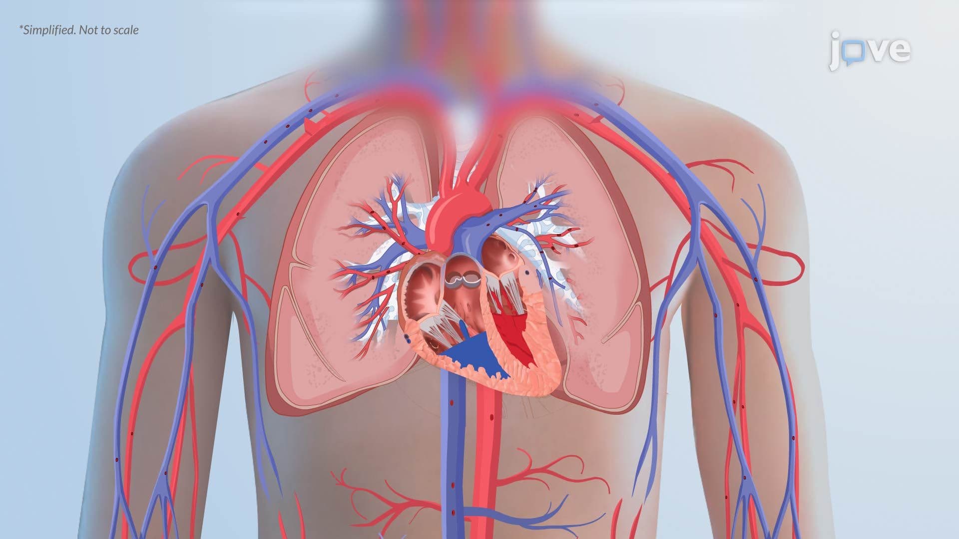

Video Summary: Pathophysiology in Pulmonary Edema Ii
Every year, nearly one million Americans are hospitalized for pulmonary edema, yet many don't know what's actually happening inside their lungs. Pathophysiology in Pulmonary Edema II breaks down the two major mechanisms, cardiogenic and noncardiogenic, that flood the alveoli and cripple gas exchange. From heart failure patients in US emergency rooms to ARDS survivors in ICUs, this concept has real-world stakes. Watch the full video on JoVE Coach to master this concept with expert-led visuals and step-by-step explanations.
Pulmonary edema is not a single disease, it's a dangerous endpoint that multiple conditions can trigger. Understanding its pathophysiology means understanding the forces that normally keep the lungs dry and what happens when those forces break down. In Pathophysiology in Pulmonary Edema II, the focus sharpens on two fundamentally different mechanisms: one driven by pressure, the other by permeability. Mastering both is essential for anyone studying respiratory physiology, whether in an AP Biology class, a college anatomy and physiology course, or while preparing for the MCAT or USMLE Step 1.
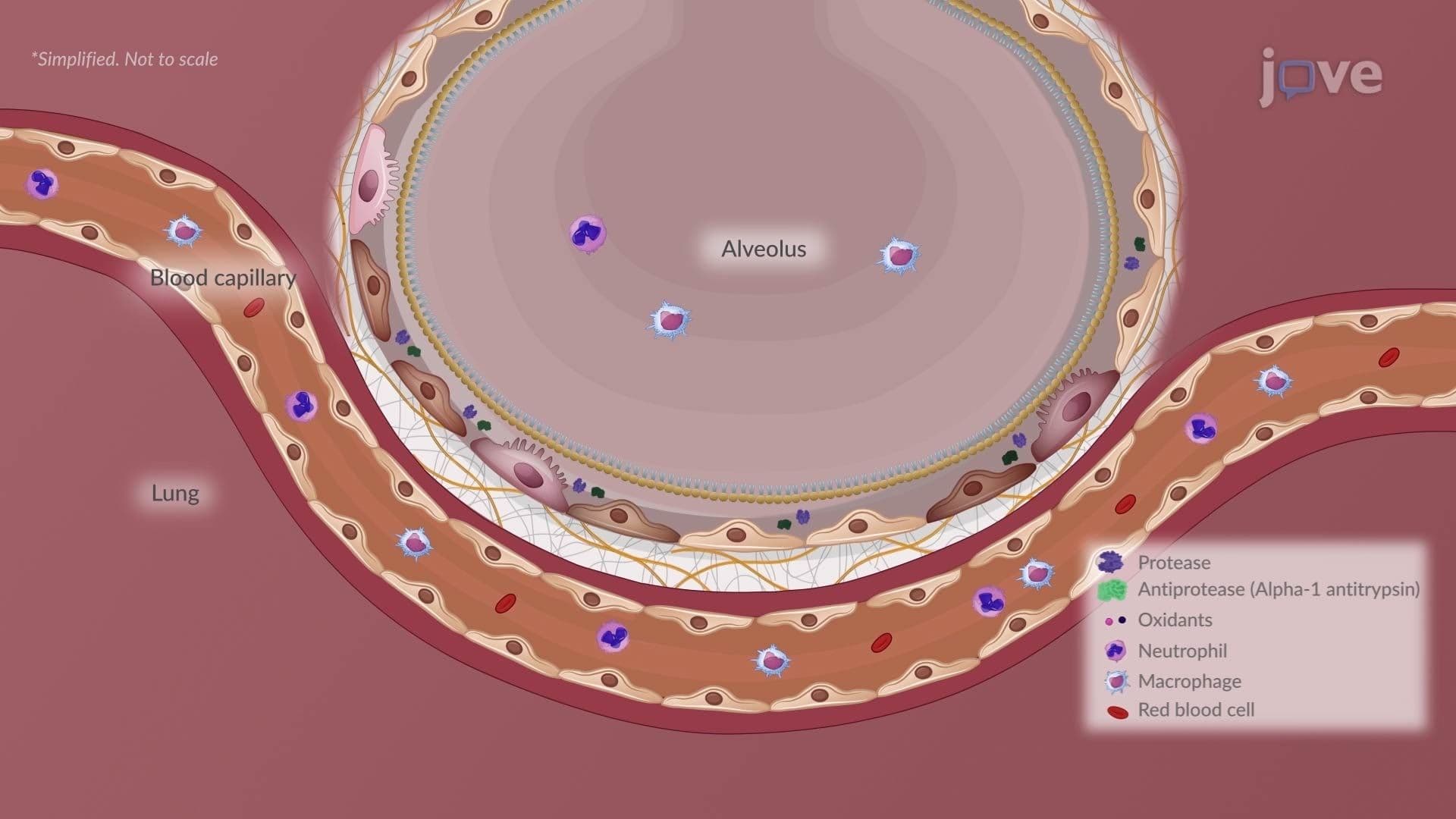
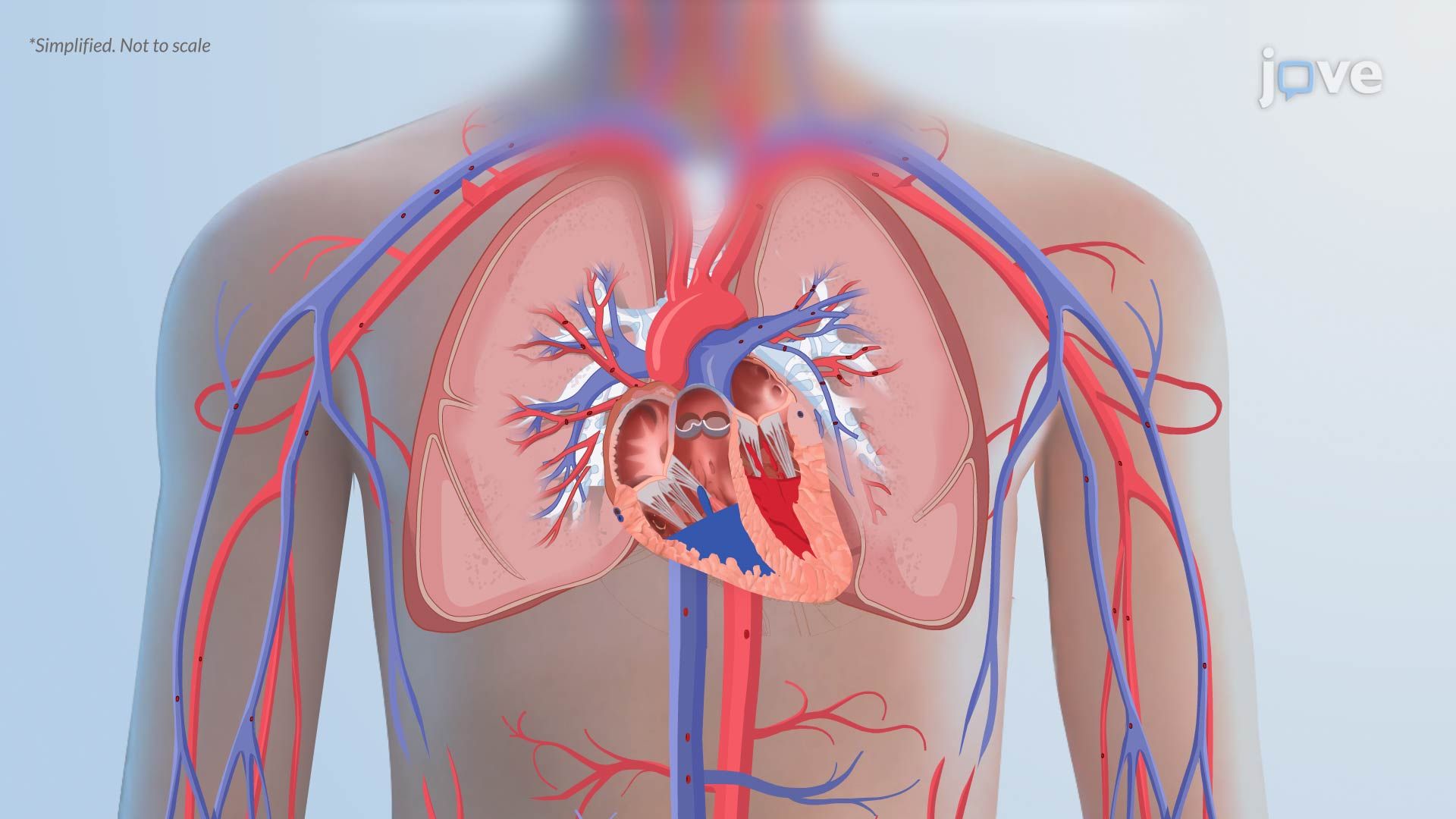
In cardiogenic pulmonary edema, the problem starts with a failing left ventricle. When the left ventricle cannot pump blood efficiently, as occurs in congestive heart failure (CHF), one of the most common hospital diagnoses in the US, blood backs up into the pulmonary circulation. This raises hydrostatic pressure inside the pulmonary capillaries beyond what the surrounding tissue can handle. According to Starling forces, fluid movement across capillary walls depends on the balance between hydrostatic pressure pushing fluid out and oncotic pressure pulling it back in. When hydrostatic pressure wins, fluid shifts into the interstitial space and eventually floods the alveoli. In American ICUs, this presentation, often accompanied by frothy pink sputum and bilateral "wet" lung sounds, is a classic clinical emergency.
Noncardiogenic edema follows a completely different path. Here, the capillary walls themselves become abnormally leaky, a hallmark of Acute Respiratory Distress Syndrome (ARDS). ARDS can be triggered by sepsis, toxic inhalation (such as smoke inhalation in wildfire victims, an increasingly relevant scenario in the US West), severe pneumonia, or trauma. In this setting, inflammatory mediators damage the capillary endothelium, allowing protein-rich fluid to escape into the alveoli. Because proteins carry osmotic pulling power, they draw even more water into the alveolar space, compounding the damage. This protein-rich flooding is what distinguishes noncardiogenic from cardiogenic edema and is a clinically critical distinction tested on both the MCAT and USMLE exams.
Regardless of the cause, once fluid enters the alveoli, it interferes with pulmonary surfactant, the detergent-like substance produced by Type II pneumocytes that normally reduces surface tension and keeps alveoli open. Surfactant dysfunction increases surface tension dramatically, which causes alveoli to collapse (atelectasis) and reduces lung compliance. Reduced compliance means the respiratory muscles must work much harder to inflate the lungs, explaining the severe dyspnea patients experience. This is mechanistically similar to what occurs in restrictive lung diseases, where stiff lungs require greater inspiratory effort. Students studying how asthma is managed or what causes respiratory system disorders will find that surfactant and compliance are recurring themes across multiple pulmonary conditions.
As alveolar flooding progresses, two compounding problems devastate gas exchange. First, the increased distance between capillary blood and alveolar air, because fluid fills the space, slows oxygen diffusion. Second, ventilation-perfusion (V/Q) mismatch develops: some lung units receive blood flow but little ventilation (due to collapsed or flooded alveoli), meaning deoxygenated blood passes through without picking up oxygen. The result is hypoxemia, dangerously low blood oxygen, and eventually hypercapnia as CO2 builds up. This is the same physiological mechanism that explains poor gas exchange in conditions like COPD, pulmonary embolism, and severe pneumonia, making Pathophysiology in Pulmonary Edema II a conceptual bridge across the entire spectrum of respiratory disease. On AP Biology and college physiology midterms, V/Q mismatch is a high-yield topic that frequently appears in free-response and multiple-choice questions alike.
Related Micro-courses