Loading subject page…

1,429 views

Video Summary: What Is Colonisation of Pathogens
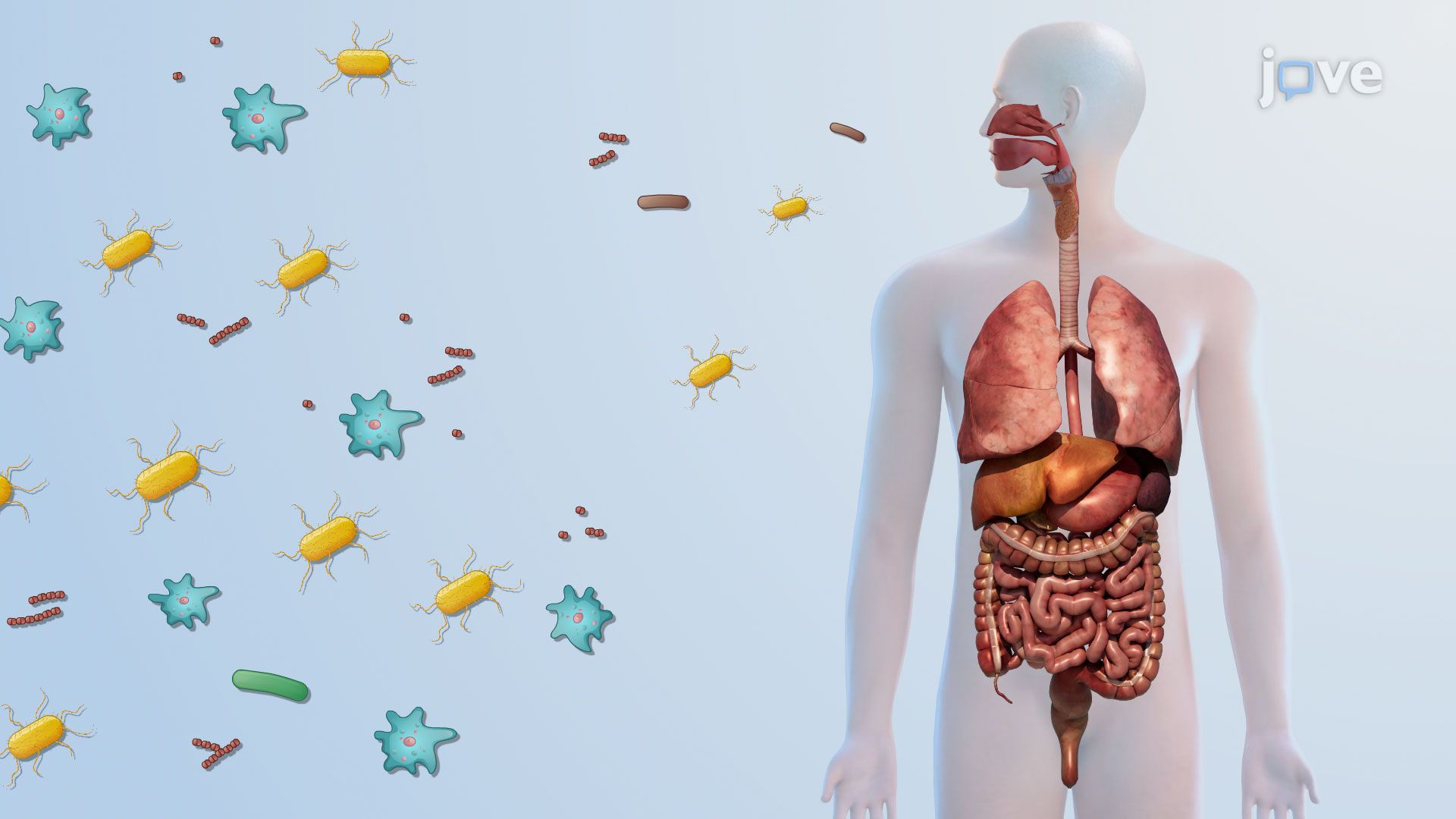
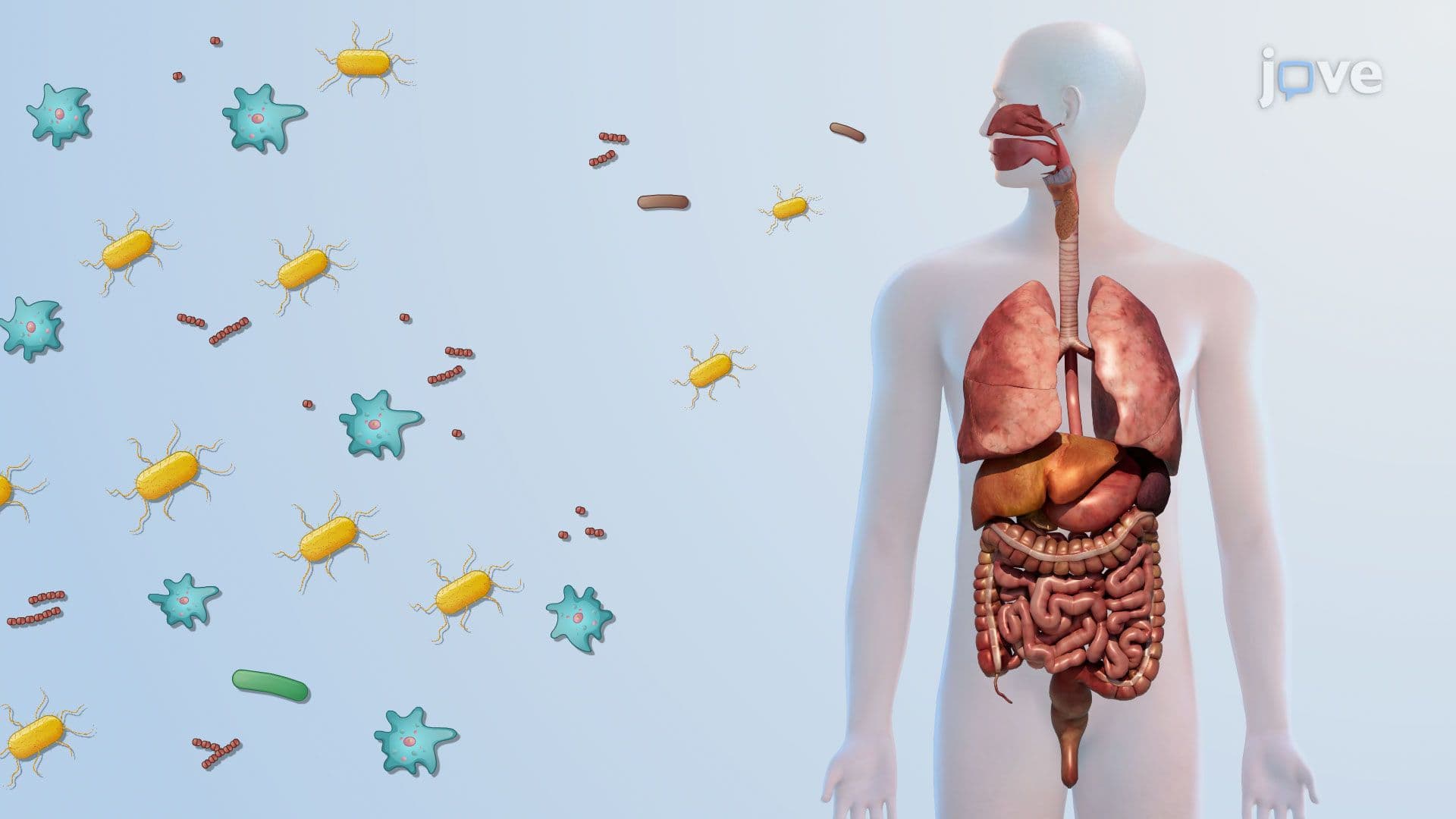
Did you know that dangerous bacteria can live inside your body for days before you feel sick? Understanding the colonisation of pathogens reveals how microbes secretly establish themselves in host tissues. In US hospitals, *Staphylococcus aureus* routinely colonizes patients' nasal passages without causing immediate harm, until it does. This concept explains the critical difference between colonization and active infection. Watch the full video on JoVE Coach to master this concept with expert-led visuals and step-by-step explanations.
The colonisation of pathogens describes the process by which prokaryotic or eukaryotic microorganisms establish a stable presence on or within host tissues, without necessarily causing disease right away. This distinction is clinically critical: colonization becomes infection only when the pathogen triggers tissue damage or provokes a significant immune response. For AP Biology students and pre-med undergraduates alike, understanding this boundary is foundational to microbiology and immunology coursework.
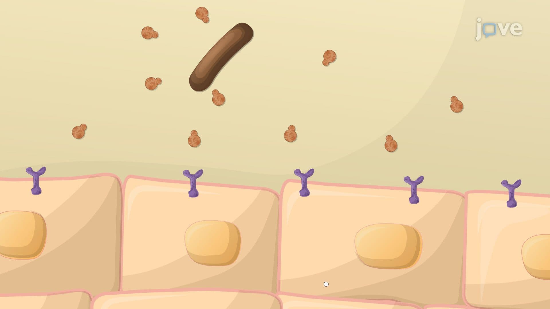
Colonization begins with physical attachment to host cells. Many bacteria deploy hair-like surface appendages called pili (or fimbriae) that grip the epithelial lining of target tissues, commonly the respiratory tract, urinary tract, or gastrointestinal mucosa. Beyond these structural anchors, pathogens express specialized surface proteins called adhesins that bind to specific receptor molecules on host cells, dramatically strengthening attachment.
A well-known US clinical example is *Escherichia coli*, whose type 1 fimbriae allow it to adhere to the epithelial cells lining the human bladder, a key first step in urinary tract infections (UTIs), one of the most common bacterial infections treated in American healthcare settings annually.
Once attached, pathogens work to establish a durable presence. One of the most powerful strategies is biofilm formation, the development of a structured, matrix-enclosed microbial community. Biofilms are notoriously resistant to both antibiotic treatment and immune clearance. In US hospitals, biofilm-forming bacteria such as *Pseudomonas aeruginosa* are a leading cause of chronic infections in patients with cystic fibrosis and those using medical implants like catheters or prosthetic heart valves.
For MCAT and USMLE test-takers, biofilm resistance is a high-yield concept frequently linked to questions about antibiotic susceptibility testing and treatment failure.
After establishing colonization, many pathogens employ sophisticated strategies to avoid destruction and spread deeper into host tissue. Two major immune evasion mechanisms stand out:
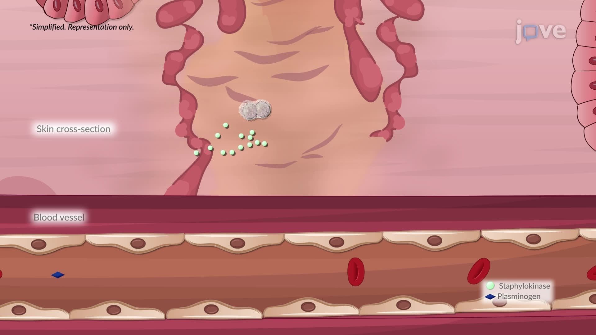
Additionally, pathogens may secrete tissue-degrading enzymes: hyaluronidase breaks down the connective tissue matrix to open pathways for deeper invasion, while coagulase (produced by *Staphylococcus aureus*) converts fibrinogen to fibrin, helping the bacterium evade phagocytosis by forming a protective clot around itself.
For high school and college students studying biology, microbiology, or health sciences, colonisation of pathogens is a recurring concept in AP Biology, college microbiology midterms, and standardized exams like the MCAT. It underpins clinical fields including infectious disease diagnostics, molecular diagnostics, and the work of clinical microbiologists who process patient specimens daily. Understanding how pathogens colonize tissue explains why proper specimen collection and transport matters, and why diagnostic microbiology techniques must detect the difference between harmless colonizers and active infectious agents.
Related Micro-courses