Loading subject page…

287 views
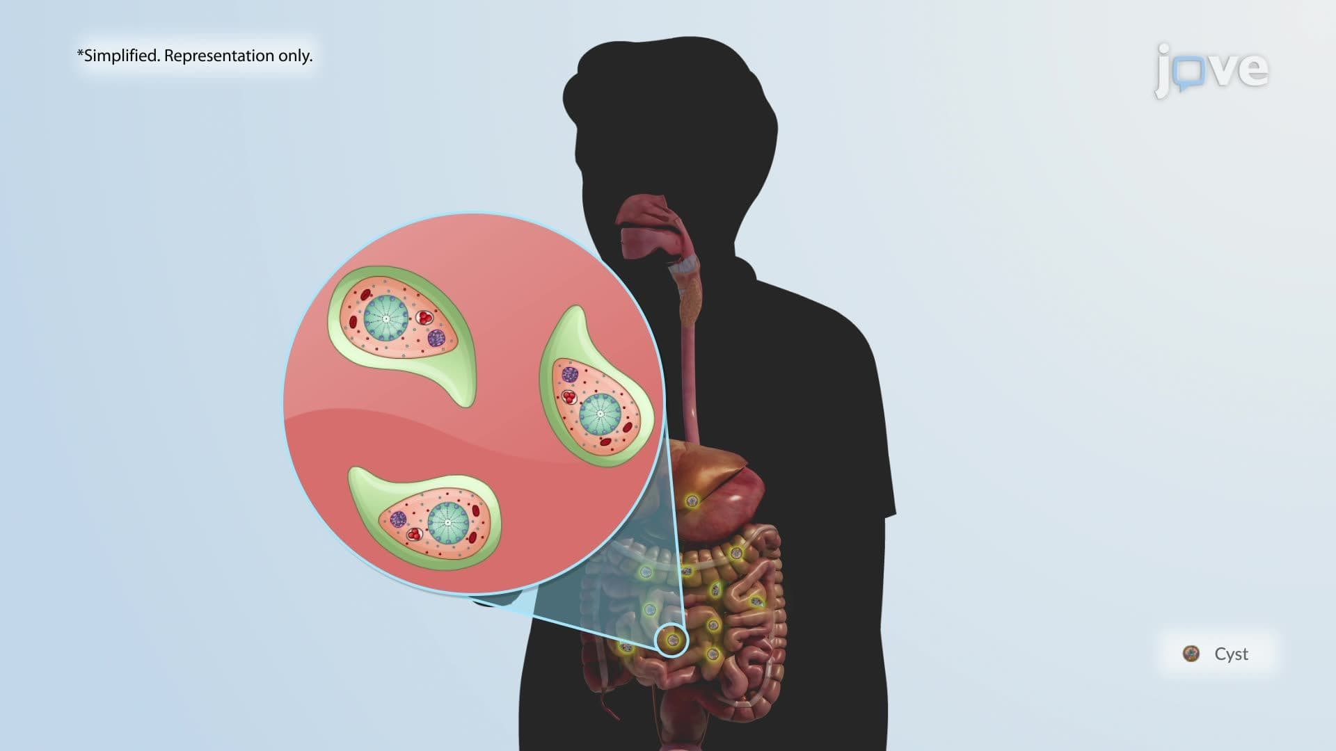

Video Summary: What Is Amebiasis
Millions of Americans travel to regions where contaminated drinking water silently spreads a dangerous gut parasite, and most never realize their risk. Amebiasis, caused by the protozoan *Entamoeba histolytica*, spreads through the fecal-oral route, triggering intestinal inflammation that can escalate to severe dysentery. Returned travelers to the US frequently present with this condition in emergency rooms. Watch the full video on JoVE Coach to master this concept with expert-led visuals and step-by-step explanations.
Amebiasis is a gastrointestinal infection caused by *Entamoeba histolytica*, a microscopic protozoan parasite capable of invading the lining of the large intestine. While it is most prevalent in regions with poor sanitation infrastructure, amebiasis is clinically relevant in the United States, particularly among international travelers, immunocompromised individuals, and residents of communities with limited access to clean water. According to the CDC, *E. histolytica* infects tens of millions of people worldwide each year, making it one of the leading parasitic causes of death globally. Understanding amebiasis builds a strong foundation in parasitology and is directly applicable to college-level microbiology and pathophysiology courses.
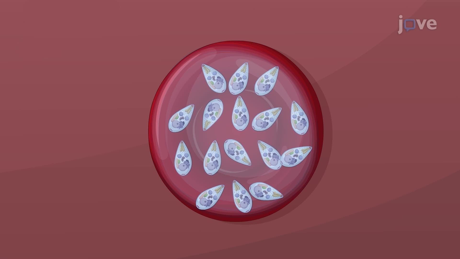
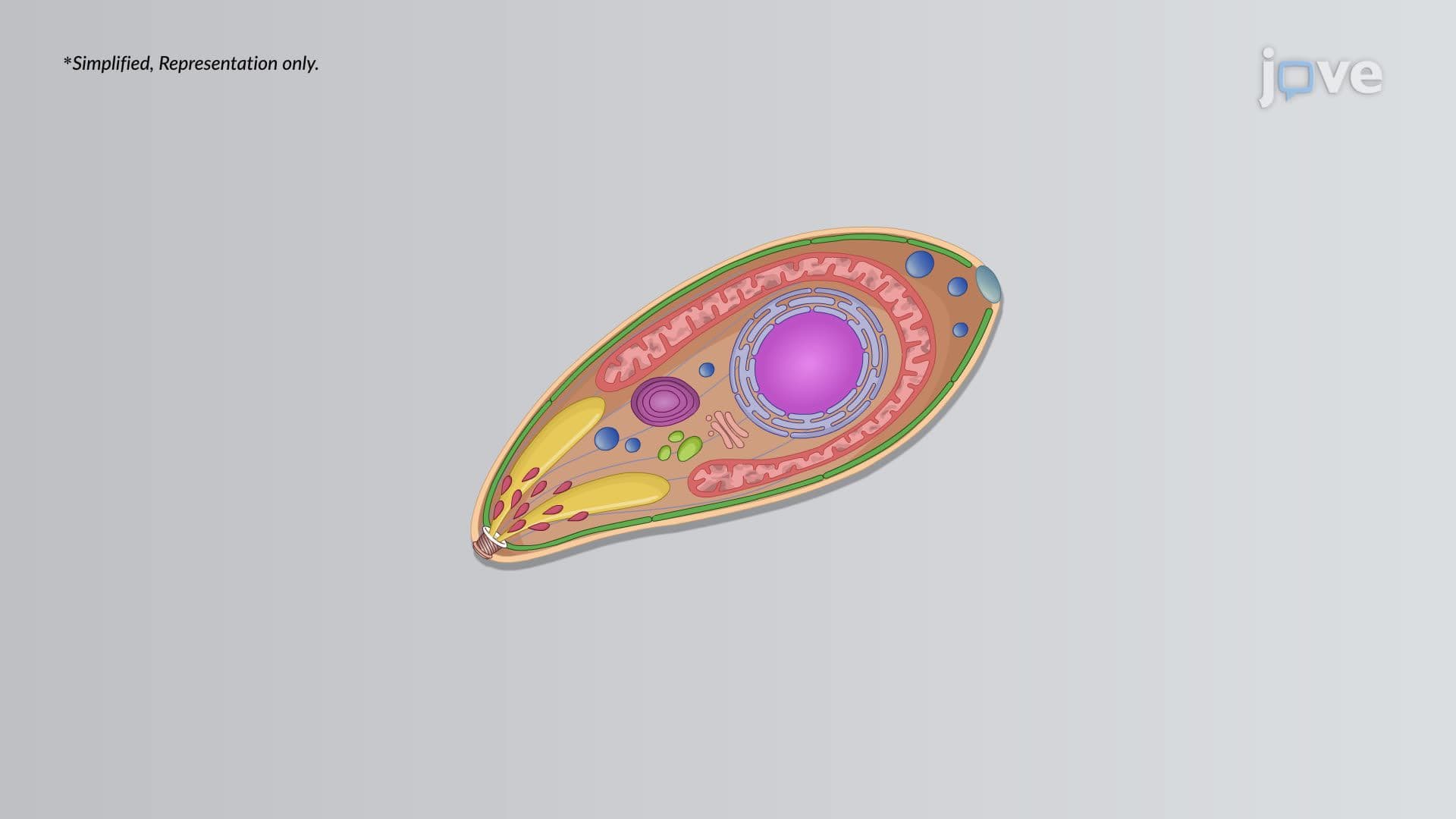
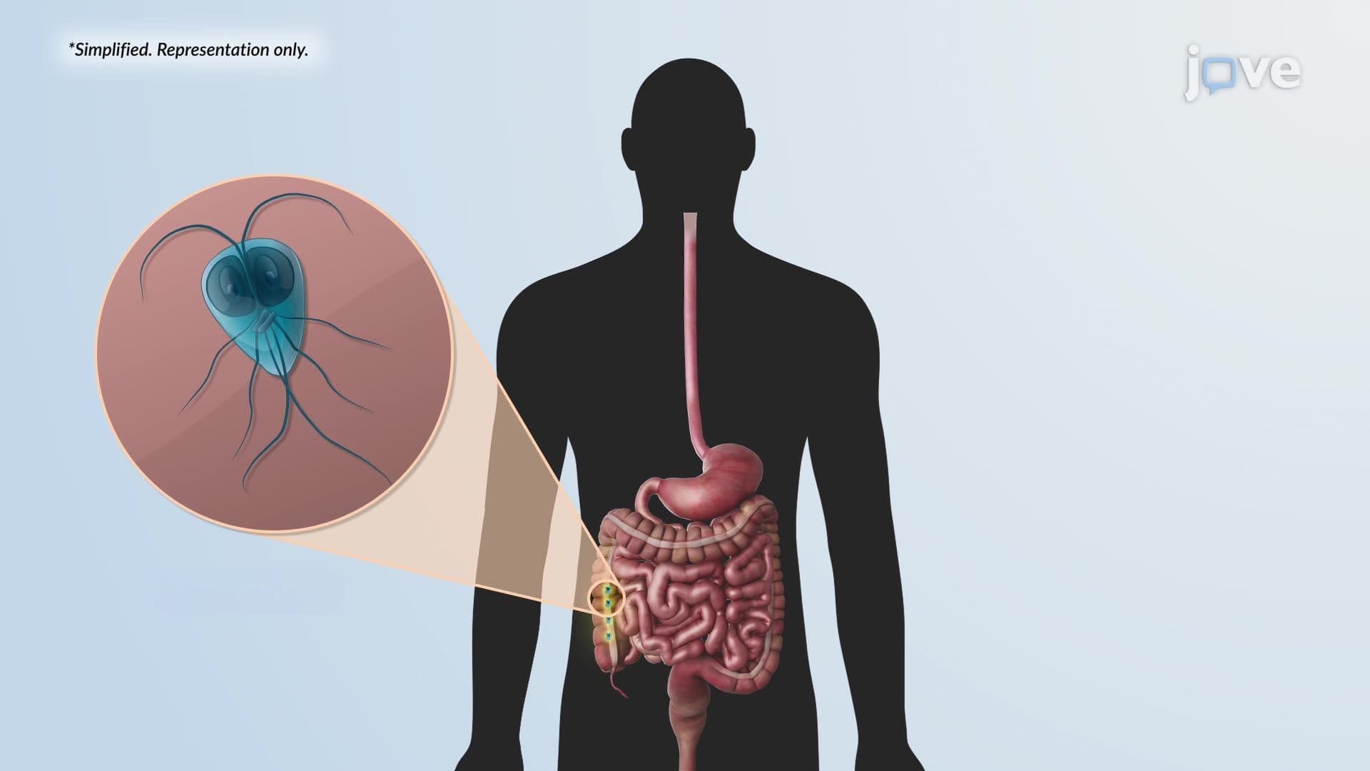
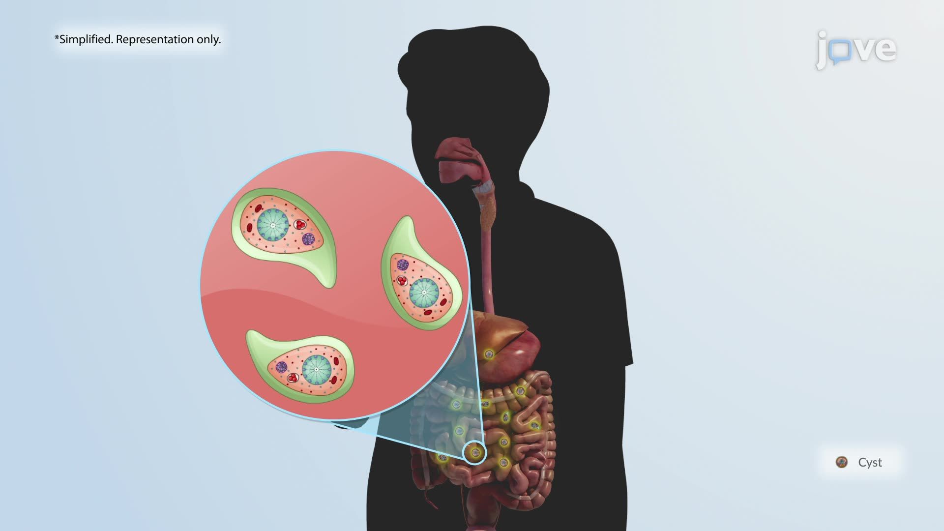
The life cycle of *Entamoeba histolytica* hinges on two key forms: the hardy, dormant cyst and the active, invasive trophozoite. Infection begins when a person swallows cysts, typically through contaminated drinking water, unwashed produce, or food handled by an infected individual. This fecal-oral transmission route is a cornerstone concept in parasitology and frequently appears on AP Biology exams and college microbiology midterms. Once ingested, the low pH of the stomach is bypassed because cysts are acid-resistant. They travel to the small intestine, where they excyst and transform into trophozoites, the metabolically active form responsible for disease.
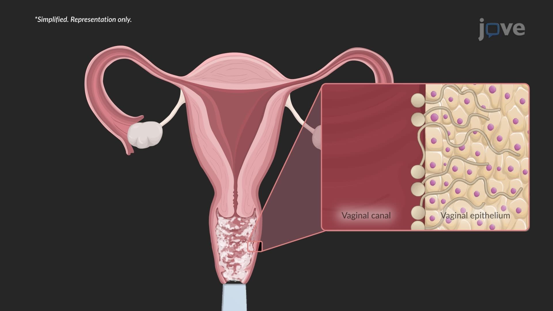
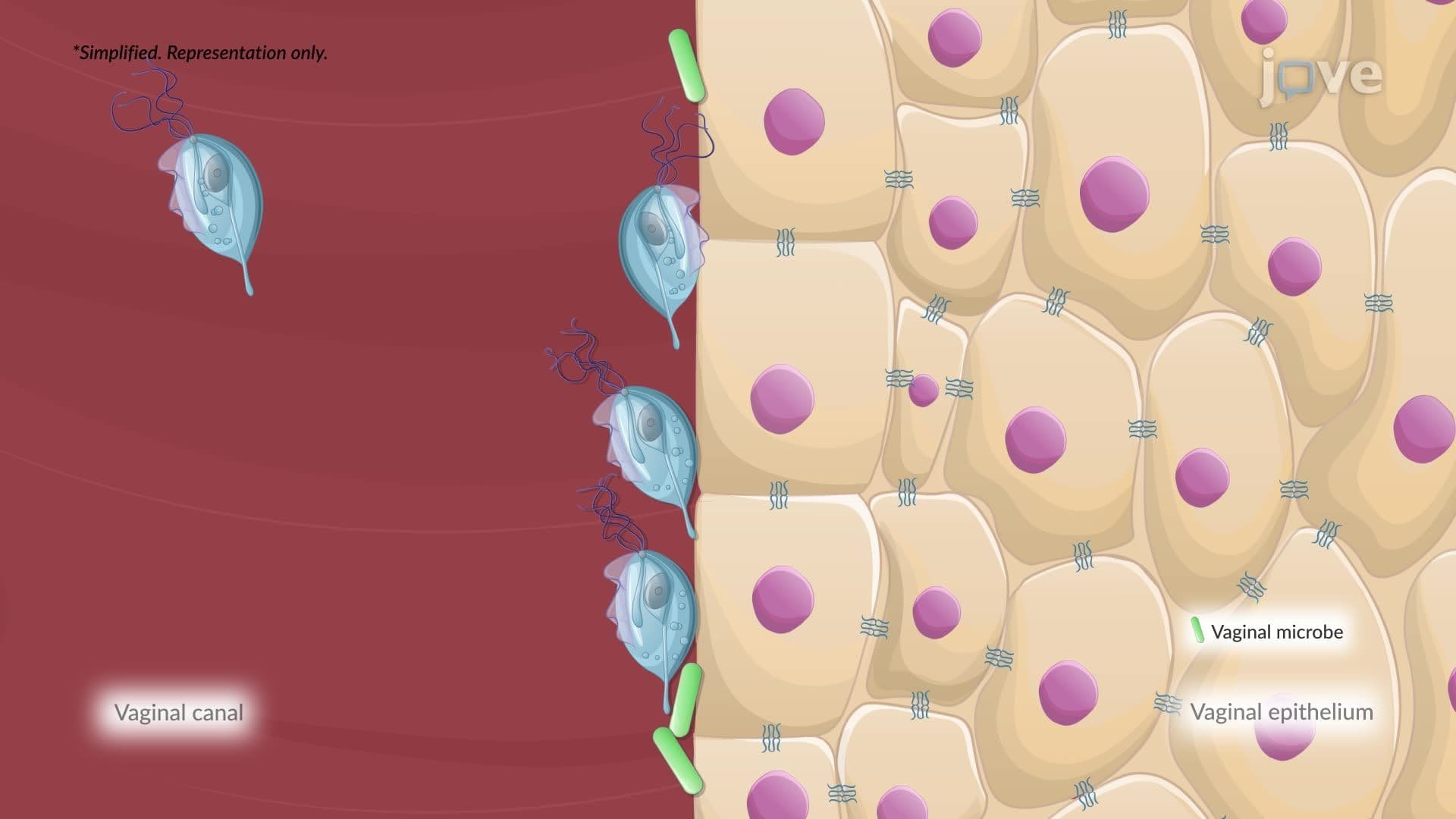
Once trophozoites reach the large intestine, they attach to colonic epithelial cells using a specialized surface molecule called galactose-N-acetylgalactosamine (Gal/GalNAc) lectin. This molecular "handshake" is what distinguishes *E. histolytica* from its non-pathogenic relative, *Entamoeba dispar*. After adhesion, trophozoites kill host cells through two mechanisms: cytolysis (direct membrane disruption) and apoptosis (programmed cell death). This dual attack damages the mucosal lining and triggers an inflammatory cascade. Injured epithelial cells release interleukin-1α and a precursor form of interleukin-1β. That precursor is then cleaved into its active form, which activates NF-κB, a critical transcription factor, in neighboring cells. NF-κB drives the production of additional pro-inflammatory cytokines that recruit neutrophils and macrophages to the infection site.
The immune response to amebiasis is a double-edged sword. Neutrophils and macrophages arrive to eliminate the invader, but trophozoites can directly destroy neutrophils, releasing additional damaging mediators that worsen mucosal injury. Activated macrophages then release tumor necrosis factor alpha (TNF-α), a powerful cytokine that amplifies inflammation. The cumulative result is mucosal ulceration, tissue necrosis, and the characteristic flask-shaped ulcers seen on pathology slides, a classic image featured in USMLE Step 1 and MCAT preparation materials. Clinically, this manifests as abdominal cramping, profuse diarrhea, and, in severe cases, bloody dysentery. Understanding this immune-pathogen interplay is essential for students in AP Biology, college immunology, and pre-health tracks preparing for standardized exams.
Related Micro-courses