Loading video…

 9 Concepts
 287 views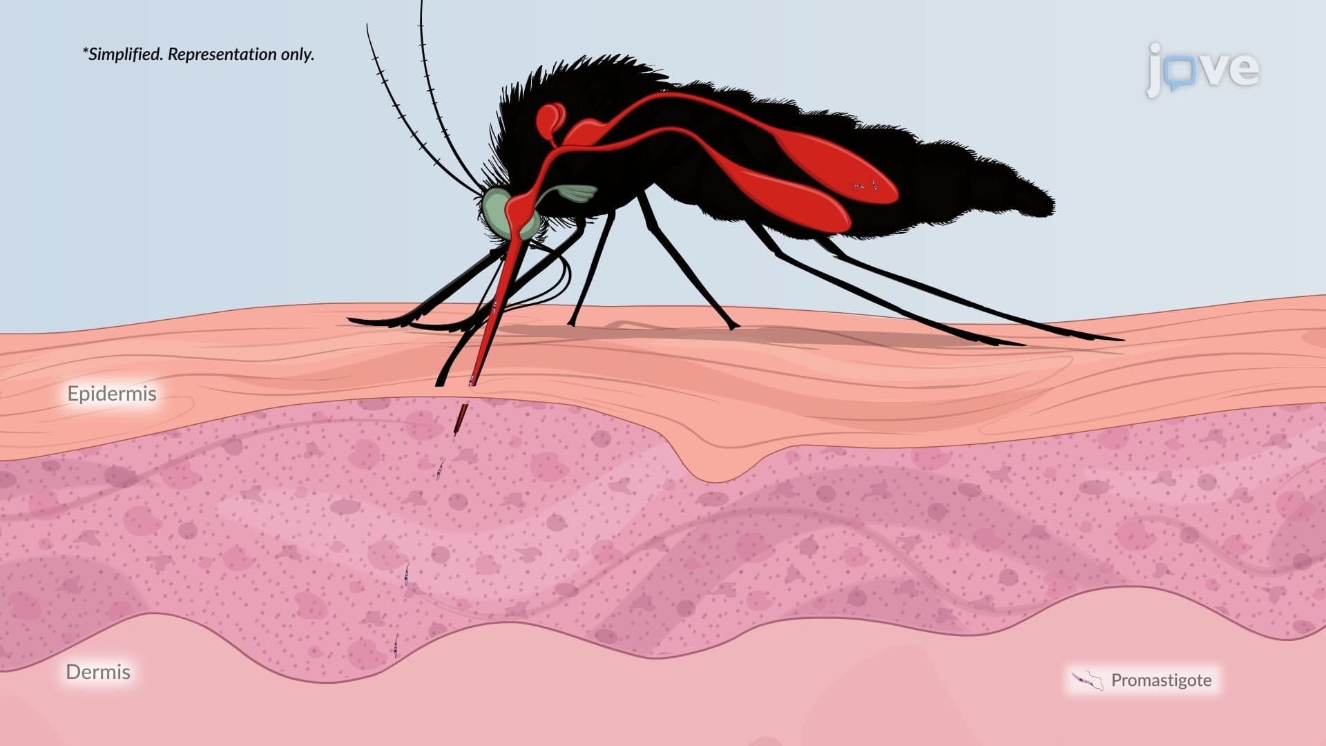

Video Summary: What Is Leishmaniasis
Did you know a tiny sandfly bite can trigger a flesh-eating ulcer, or even cause organ failure? Leishmaniasis, a parasitic disease caused by *Leishmania* protozoa, affects over a million people worldwide each year and has been documented in US military personnel returning from the Middle East. Understanding what is leishmaniasis means tracing a parasite's clever journey from sandfly to human macrophage. Watch the full video on JoVE Coach to master this concept with expert-led visuals and step-by-step explanations.
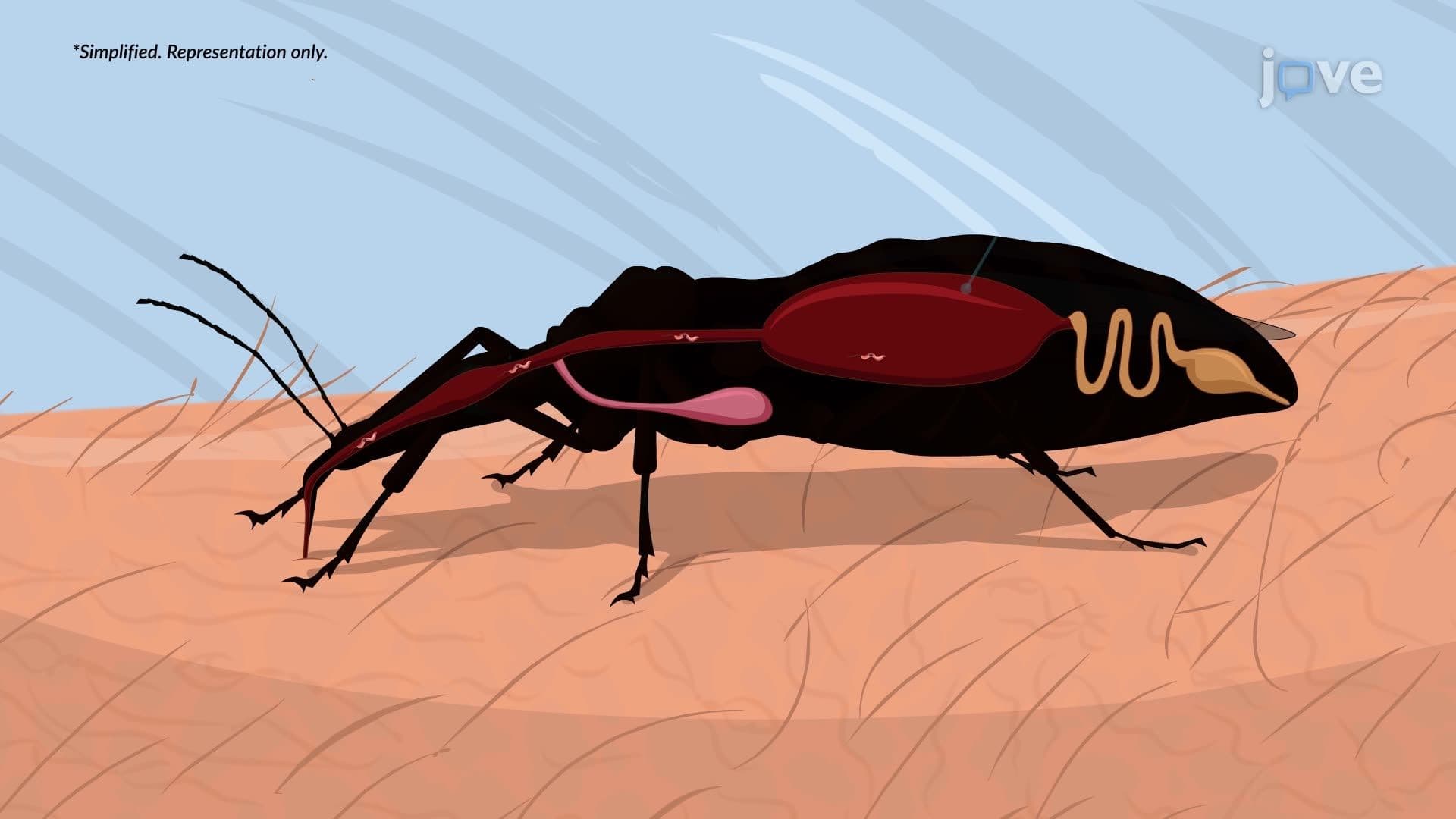
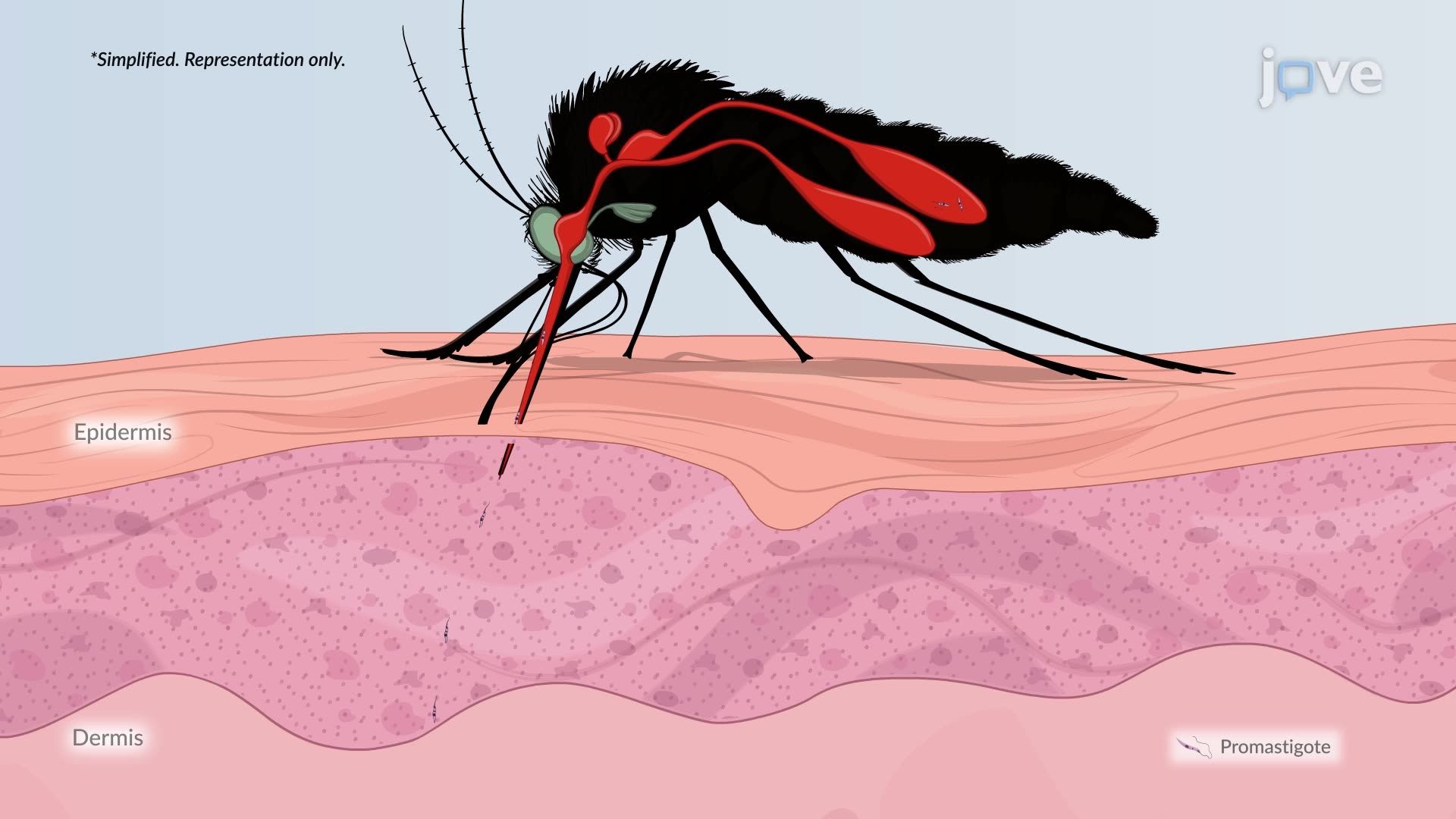
Leishmaniasis is a vector-borne protozoal disease caused by over 20 species of the genus *Leishmania*. It is transmitted exclusively through the bite of an infected female phlebotomine sandfly, a small, blood-feeding insect found in tropical, subtropical, and some temperate regions. While leishmaniasis is most common in parts of South Asia, East Africa, and Latin America, it holds real clinical relevance in the United States: the CDC has documented cases among US soldiers deployed to Iraq and Afghanistan, where cutaneous leishmaniasis caused by *L. major* and *L. tropica* has been an ongoing health concern. Understanding this disease is essential for students in AP Biology, college microbiology, and those preparing for exams like the MCAT or USMLE.
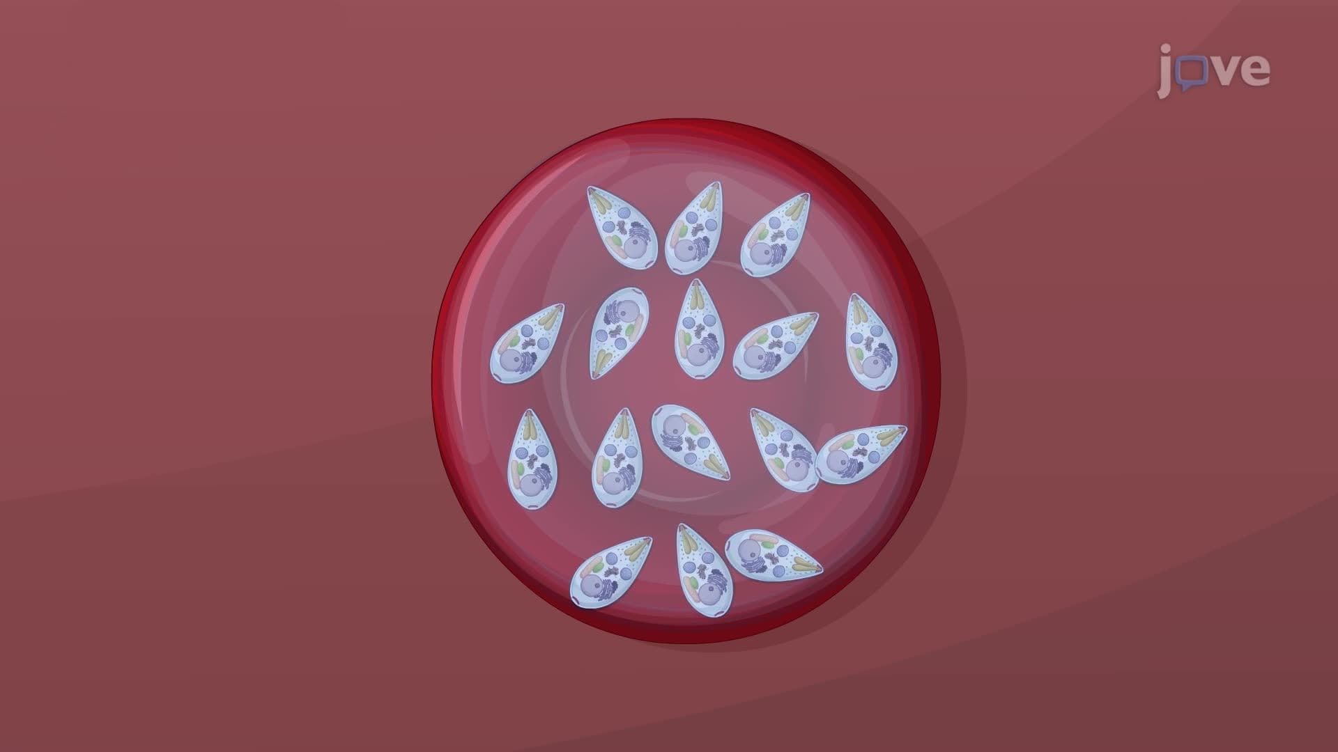
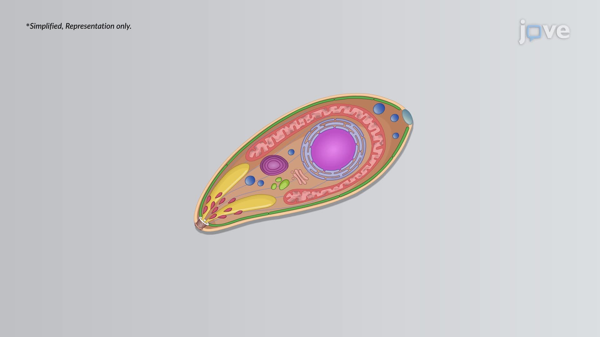
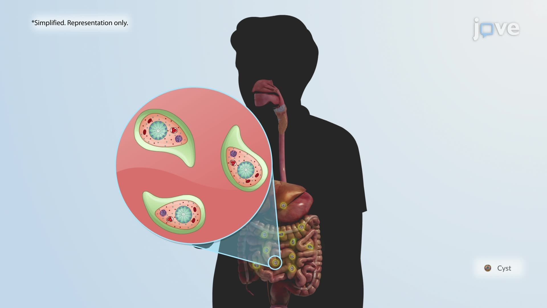
The life cycle of *Leishmania* is a classic example of a digenetic parasite, one that requires two hosts to complete development. In the animal reservoir (such as a dog or rodent), the parasite exists as an amastigote: a non-motile, intracellular form lacking an external flagellum. When a female sandfly takes a blood meal from an infected host, it ingests these amastigotes. Inside the sandfly's midgut, amastigotes transform into promastigotes, elongated, flagellated, and highly motile forms capable of replicating rapidly. These promastigotes migrate to the fly's mouthparts and are injected into a new human host during the next bite. This transformation between forms is a high-yield concept in parasitology courses and frequently appears on MCAT biology sections testing life cycle knowledge.
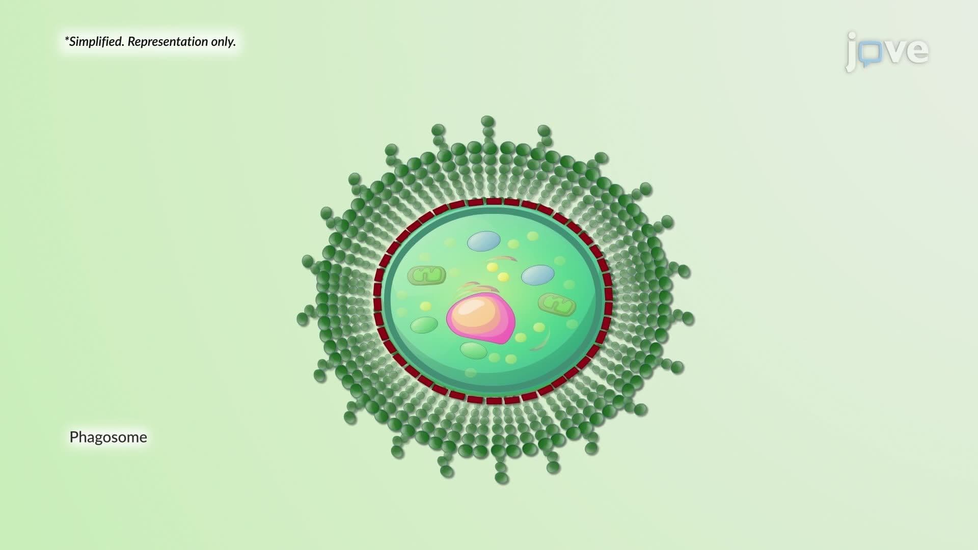
Once promastigotes enter human skin, they are quickly recognized and engulfed by skin-resident macrophages, a process called phagocytosis. Normally, phagocytosis destroys pathogens by enclosing them in phagolysosomes, organelles with acidic, enzyme-rich environments. *Leishmania*, however, has evolved remarkable acid-tolerance mechanisms that allow amastigotes to survive and replicate within these compartments. As infected macrophages eventually rupture, newly released amastigotes infect neighboring macrophages, spreading the infection locally. Clinically, this produces skin lesions that begin as painless, small papules and can progress over weeks to months into ulcers with a raised, violaceous (purplish-red) border and central epidermal breakdown, a presentation sometimes called a "volcano crater" ulcer. These lesions are a classic board exam visual clue.
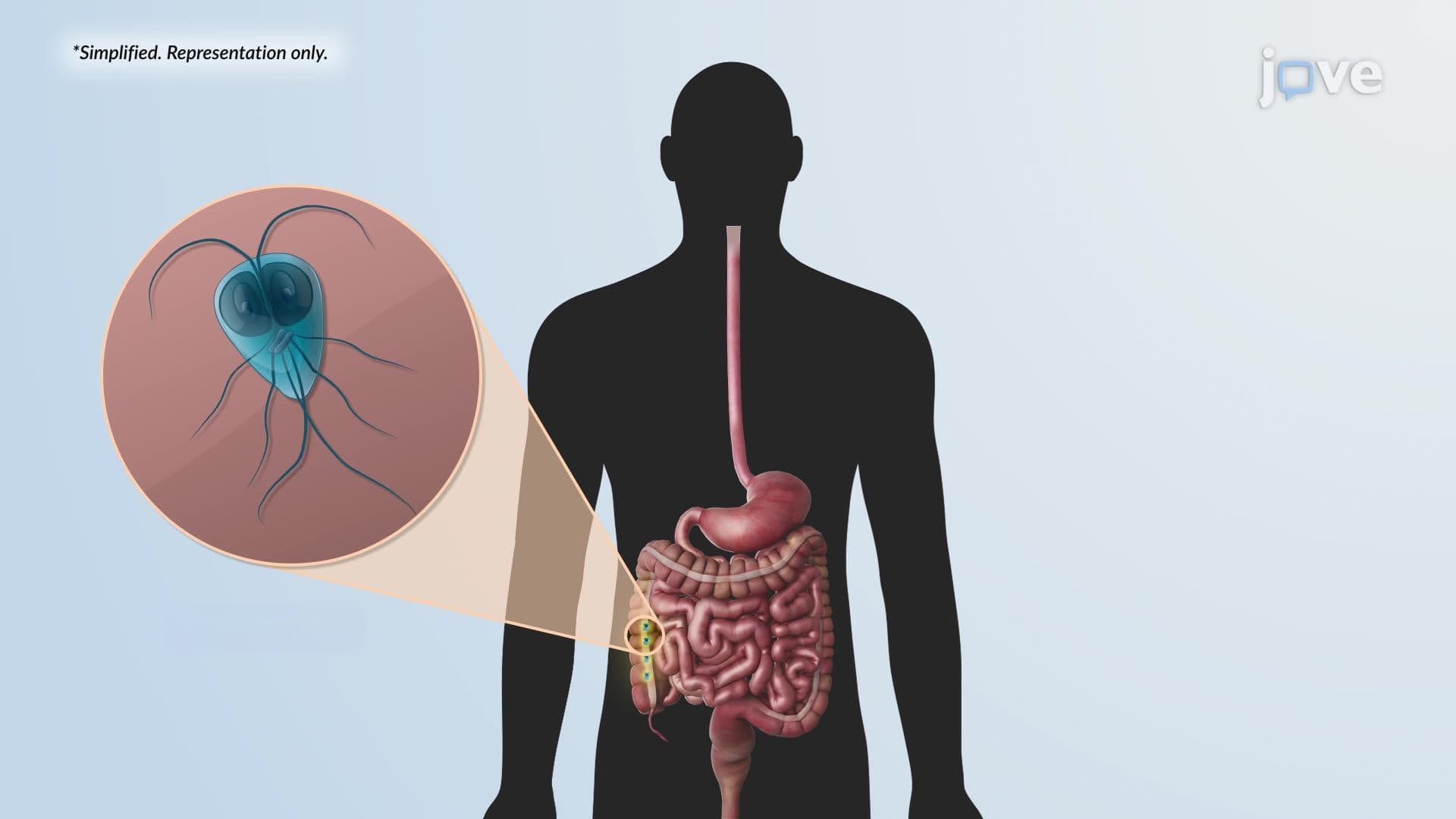
In visceral leishmaniasis, also known as kala-azar, the infection does not stay localized to the skin. Instead, amastigotes are carried through the bloodstream inside mononuclear cells (such as monocytes and macrophages) to the body's reticuloendothelial organs: the spleen, liver, bone marrow, and intestinal lymph nodes. This systemic spread causes massive splenomegaly and hepatomegaly, severe anemia (due to bone marrow involvement), fever, and dramatic weight loss. Without treatment, visceral leishmaniasis is nearly always fatal. Students in college immunology and pathology courses should connect this pattern to concepts of mononuclear phagocyte system dysfunction, a frequent topic on USMLE Step 1 and NCLEX pharmacology modules. Understanding the difference between localized cutaneous and systemic visceral forms is a critical compare-and-contrast skill tested on midterms and standardized exams alike.
Related Micro-courses