Loading subject page…

287 views
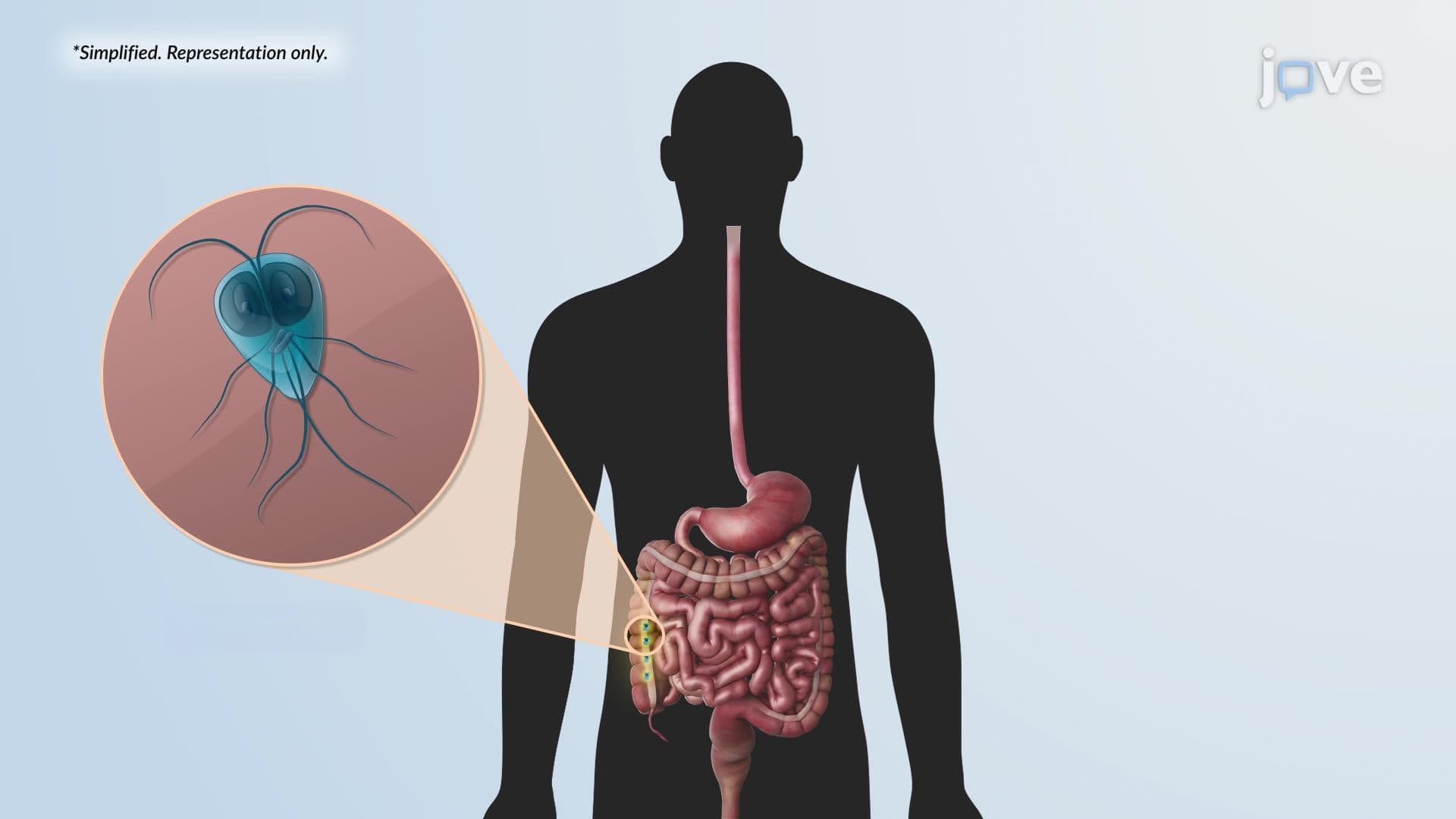

Video Summary: What Is Giardiasis
Ever wonder how a sip of contaminated lake water can leave you sick for weeks? Giardiasis, a common intestinal infection caused by the protozoan parasite *Giardia duodenalis*, is one of the most widespread waterborne illnesses in the US, affecting millions annually. This microscopic parasite hijacks your small intestine, disrupting nutrient absorption in a remarkably clever way. Watch the full video on JoVE Coach to master this concept with expert-led visuals and step-by-step explanations.
Giardiasis is an intestinal infection caused by *Giardia duodenalis* (also called *Giardia lamblia* or *Giardia intestinalis*), a microscopic protozoan parasite. It ranks among the most common parasitic gastrointestinal infections in the United States, with the CDC estimating over one million cases each year. The infection is especially prevalent among hikers and campers who drink untreated water from natural sources, earning *Giardia* the nickname "Beaver Fever." Understanding giardiasis is foundational in both parasitology and introductory microbiology courses, and it appears frequently on exams like the MCAT and AP Biology.
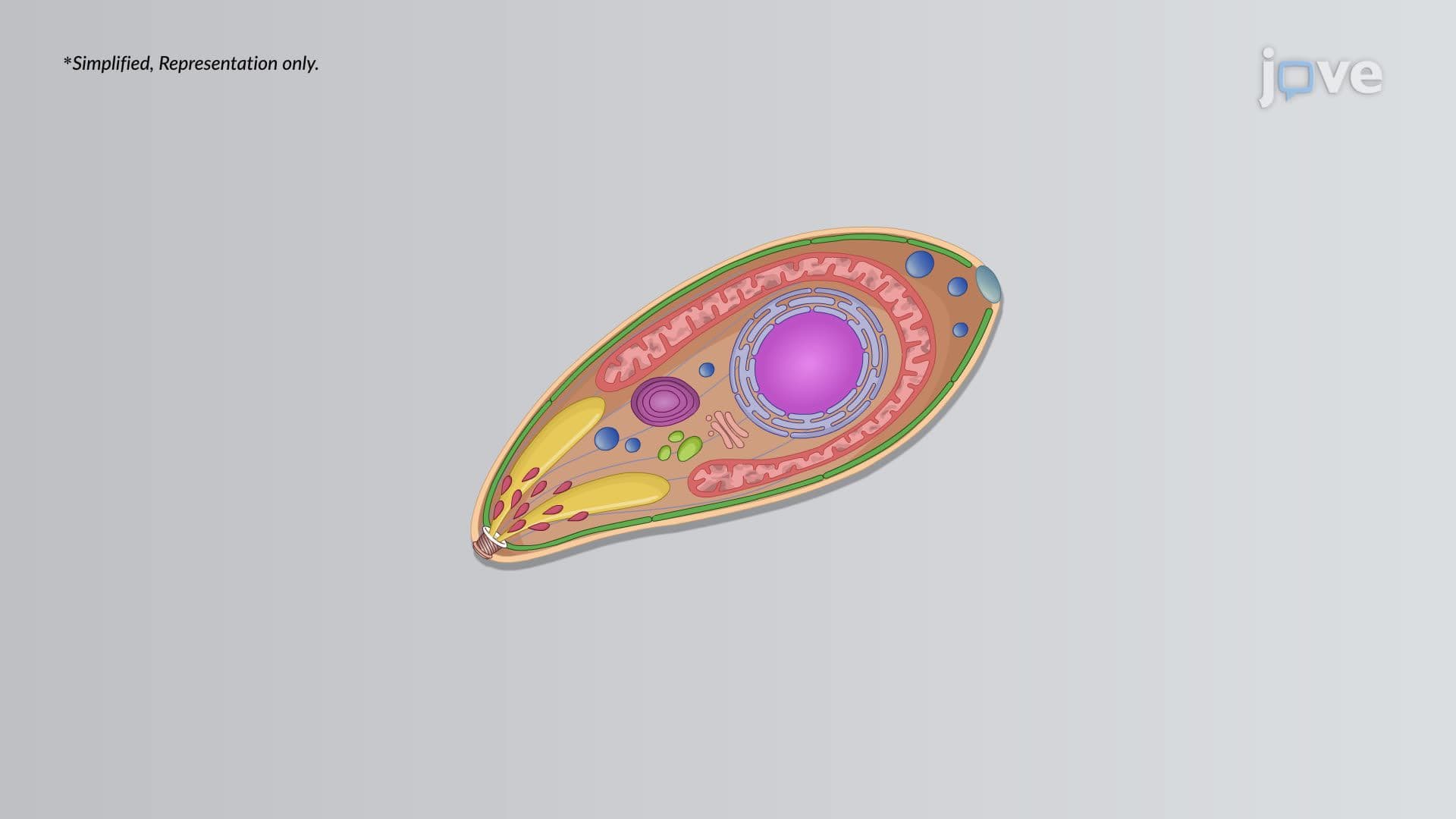
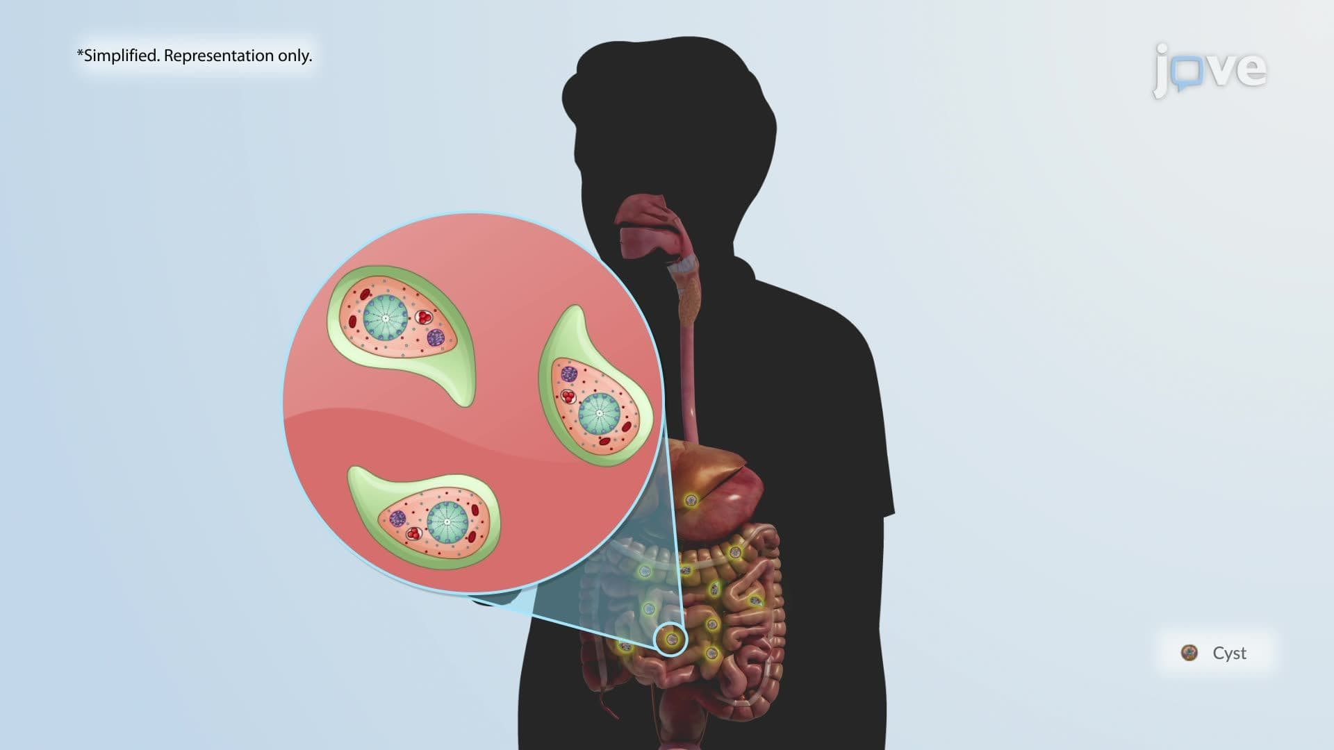
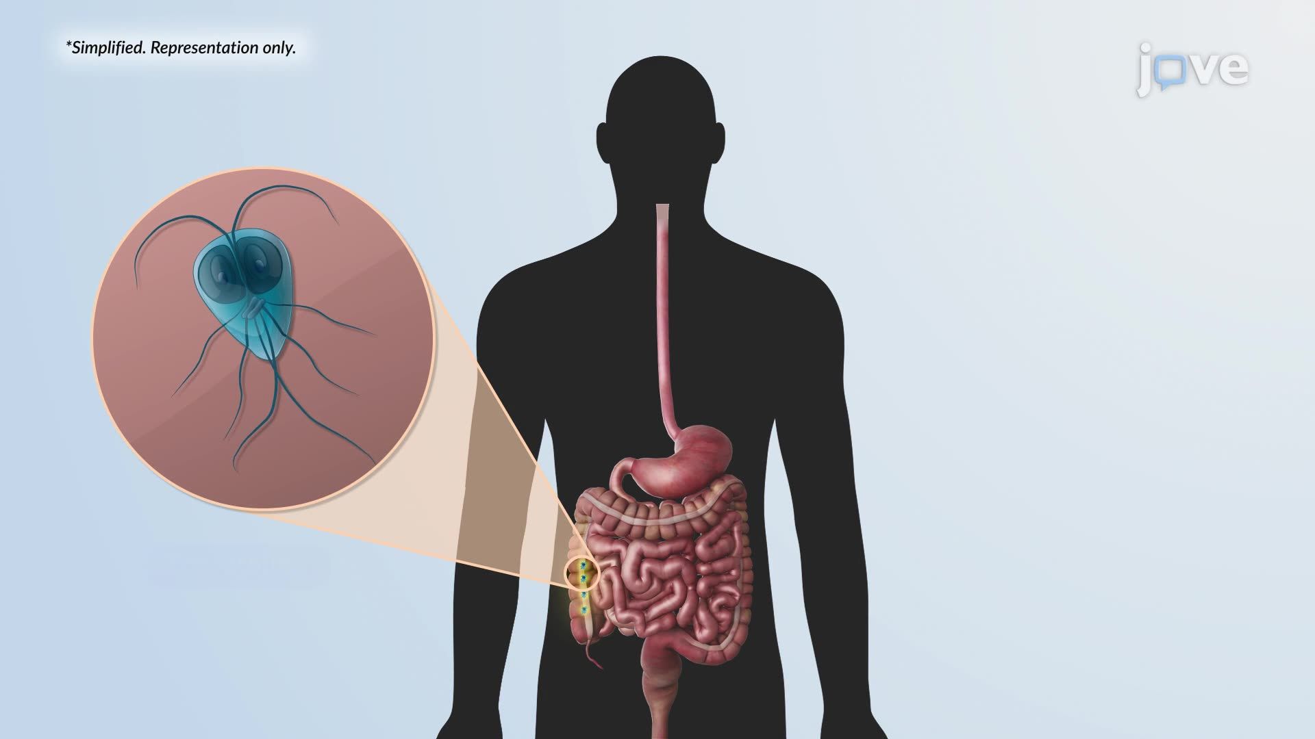
The life cycle of *Giardia* has two primary stages: the cyst and the trophozoite. The cyst is the infectious, environmentally resistant form. When a person ingests as few as 10 cysts, through contaminated drinking water, unwashed produce, or person-to-person contact, infection can occur. Inside the stomach, the low pH environment weakens the cyst wall, initiating excystation. The partially activated parasites travel to the duodenum (the first section of the small intestine), where pancreatic enzymes complete the process, releasing fully active, motile trophozoites. These pear-shaped, flagellated organisms rapidly divide and colonize the duodenal mucosa. This elegant, two-stage life cycle is a classic example of how parasites adapt to exploit the host's own digestive chemistry.
Once attached to the mucosal surface of the duodenum, trophozoites do not invade the intestinal wall, instead, they cause damage from the outside in. They secrete proteases and other parasite-derived molecules that degrade brush-border enzymes, which are critical for breaking down carbohydrates, proteins, and fats. Additionally, the physical attachment of trophozoites disrupts the microvilli, the tiny, finger-like projections that dramatically increase the surface area of the small intestine for nutrient absorption. The result is malabsorption: the body cannot efficiently absorb nutrients, leading to hallmark symptoms like fatty, foul-smelling diarrhea, bloating, gas, and weight loss. This mechanism is a strong example of a non-invasive pathogen causing significant clinical illness, a concept heavily tested in college biology and pre-health courses.
As trophozoites migrate toward the large intestine, they encounter rising pH levels and secondary bile salts, conditions that trigger encystation, the conversion back into cysts. These new cysts are excreted in feces and can survive in cold water for months, making contaminated water sources a major transmission route. In the US, outbreaks have been linked to municipal water supplies, daycare centers, and backcountry water sources in national parks like the Rocky Mountains and Appalachian Trail. This environmental resilience is why standard chlorination sometimes fails to eliminate *Giardia*, and why filtration or UV treatment is recommended for wilderness drinking water. From an exam standpoint, understanding the full cycle, ingestion → excystation → colonization → encystation → excretion, gives students a strong framework for answering questions about parasitic infection transmission, host-pathogen dynamics, and gastrointestinal disease on standardized tests.
Related Micro-courses