- Anatomy and Physiology
- The Muscular System
Micro-courses:31

The Muscular System

1. The Muscular System
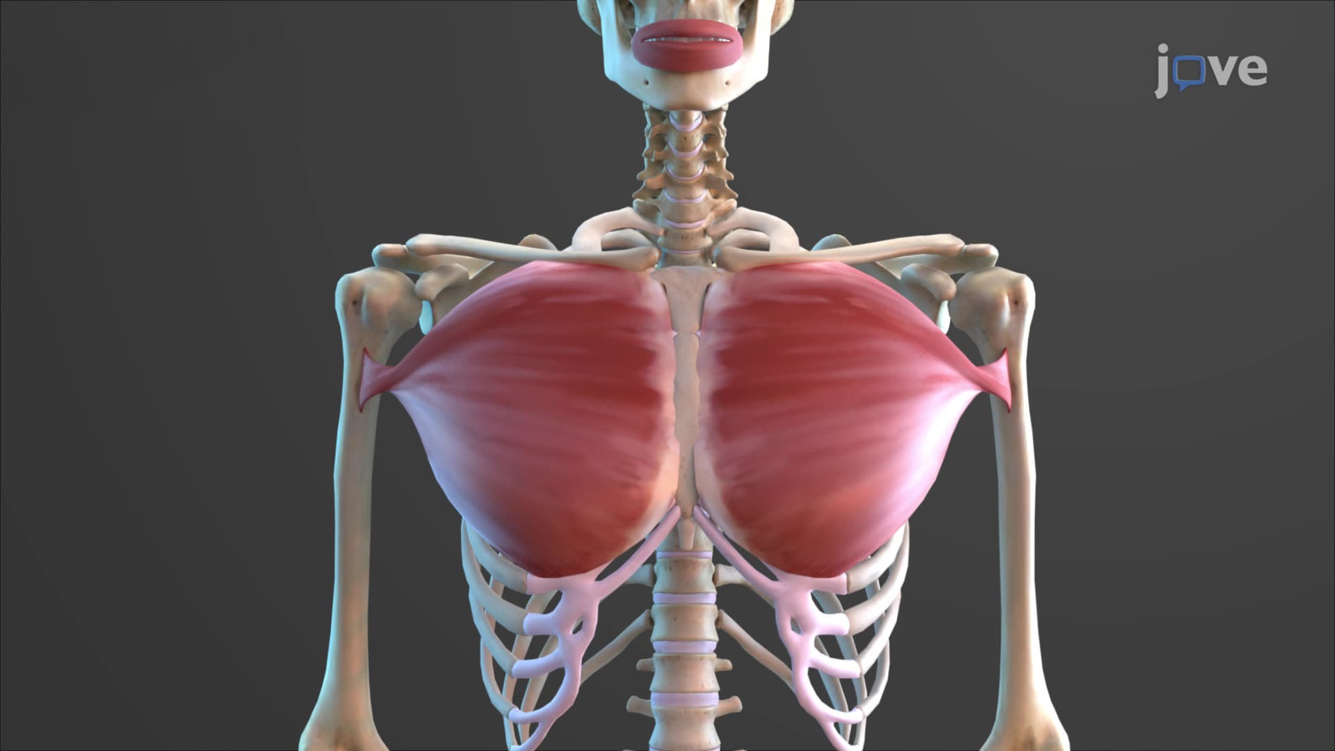

2. Fascicle Arrangement in Skeletal Muscles
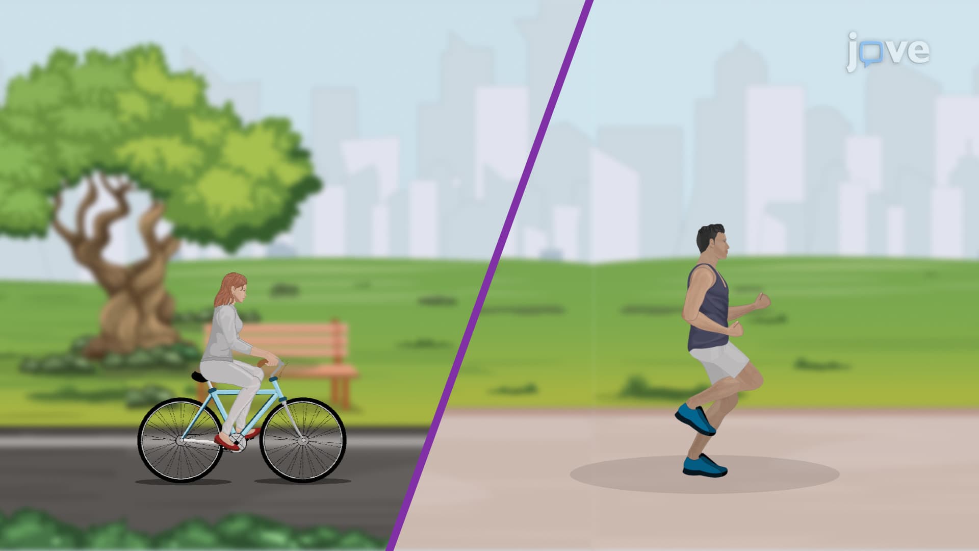
3. Muscle Coordination and Action
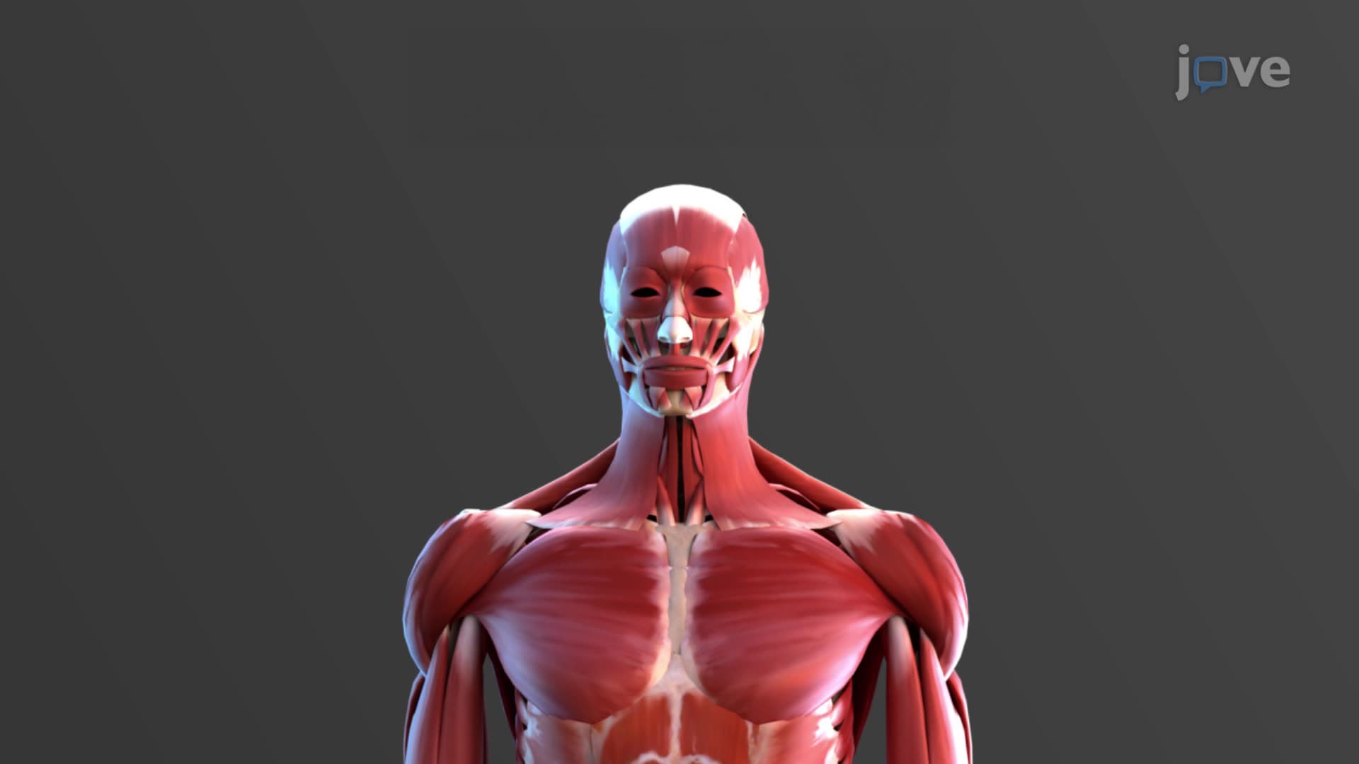
4. Naming Skeletal Muscles

5. Axial and Appendicular Muscles
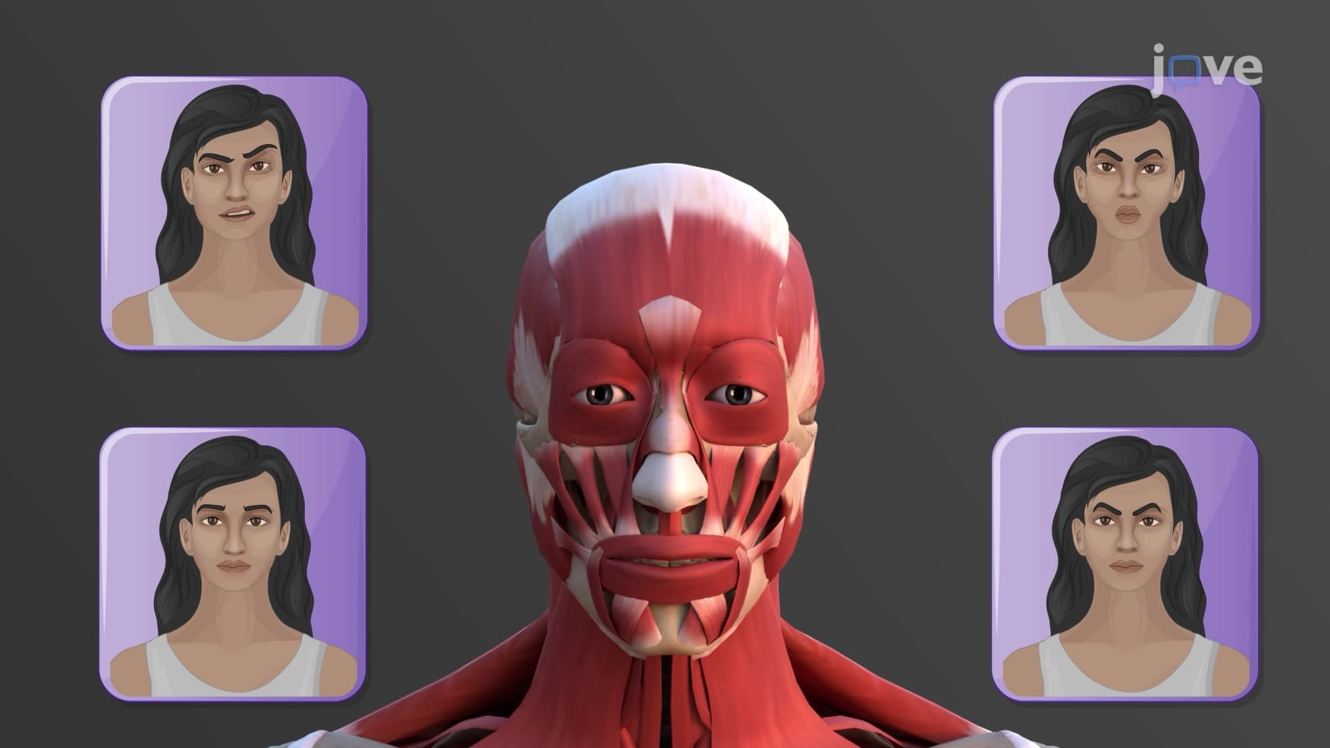
6. Muscles for Facial Expressions
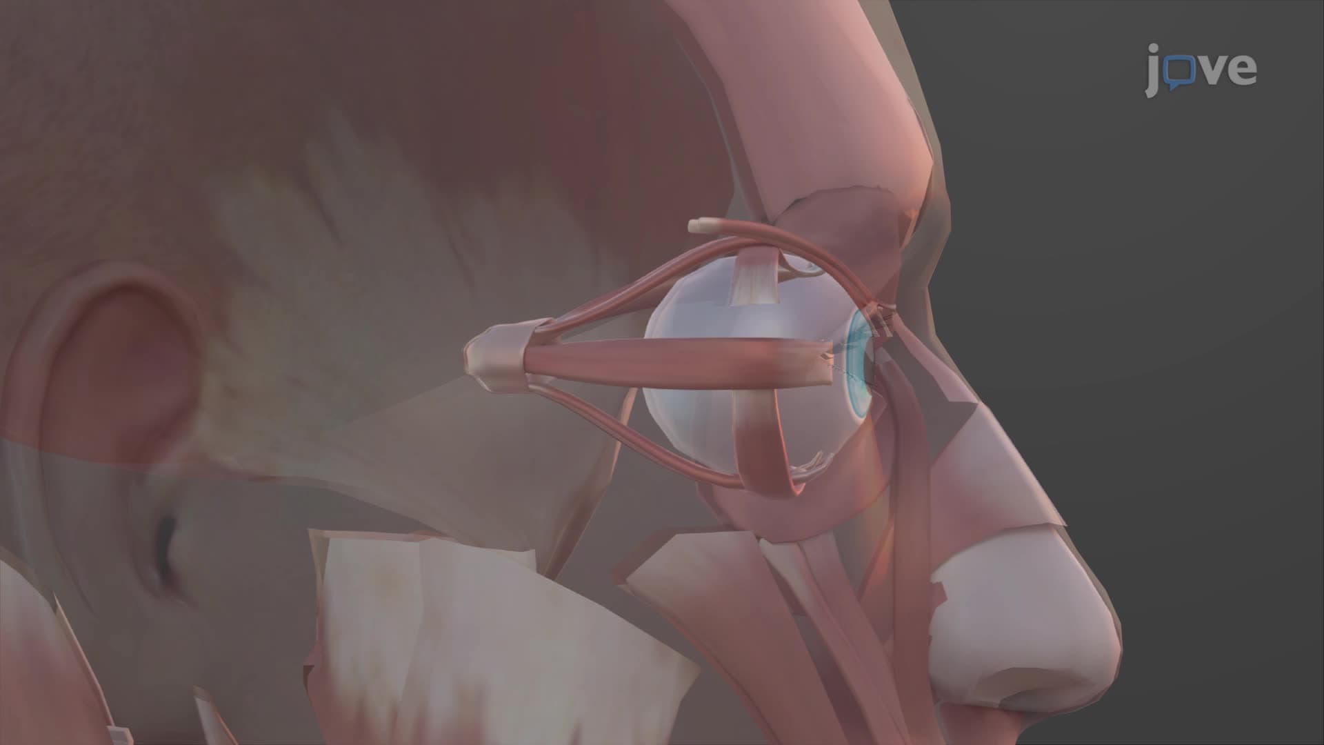
7. Muscles of the Eye
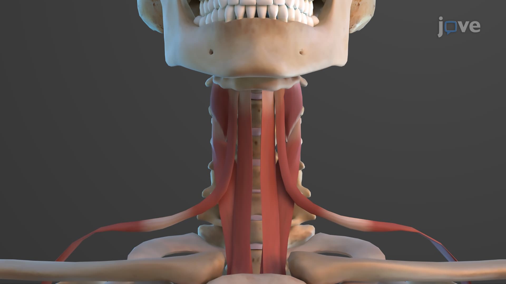
8. Muscles of the Anterior Neck
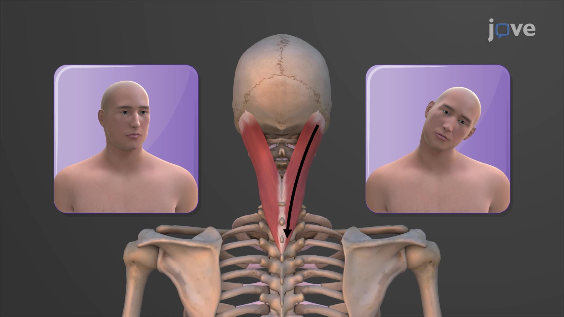
9. Muscles that Move the Head
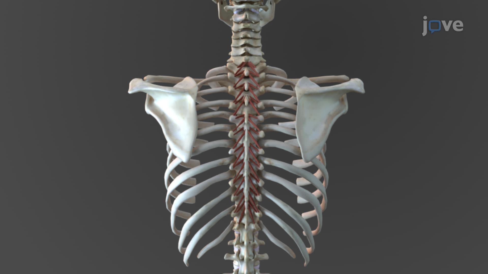
10. Muscles of the Vertebral Column
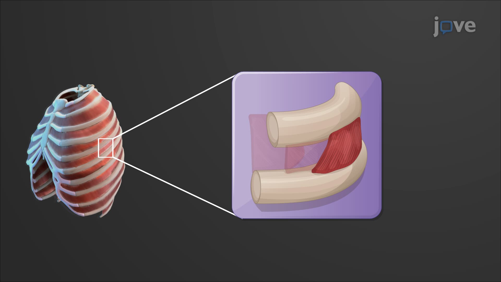
11. Muscles of the Thorax
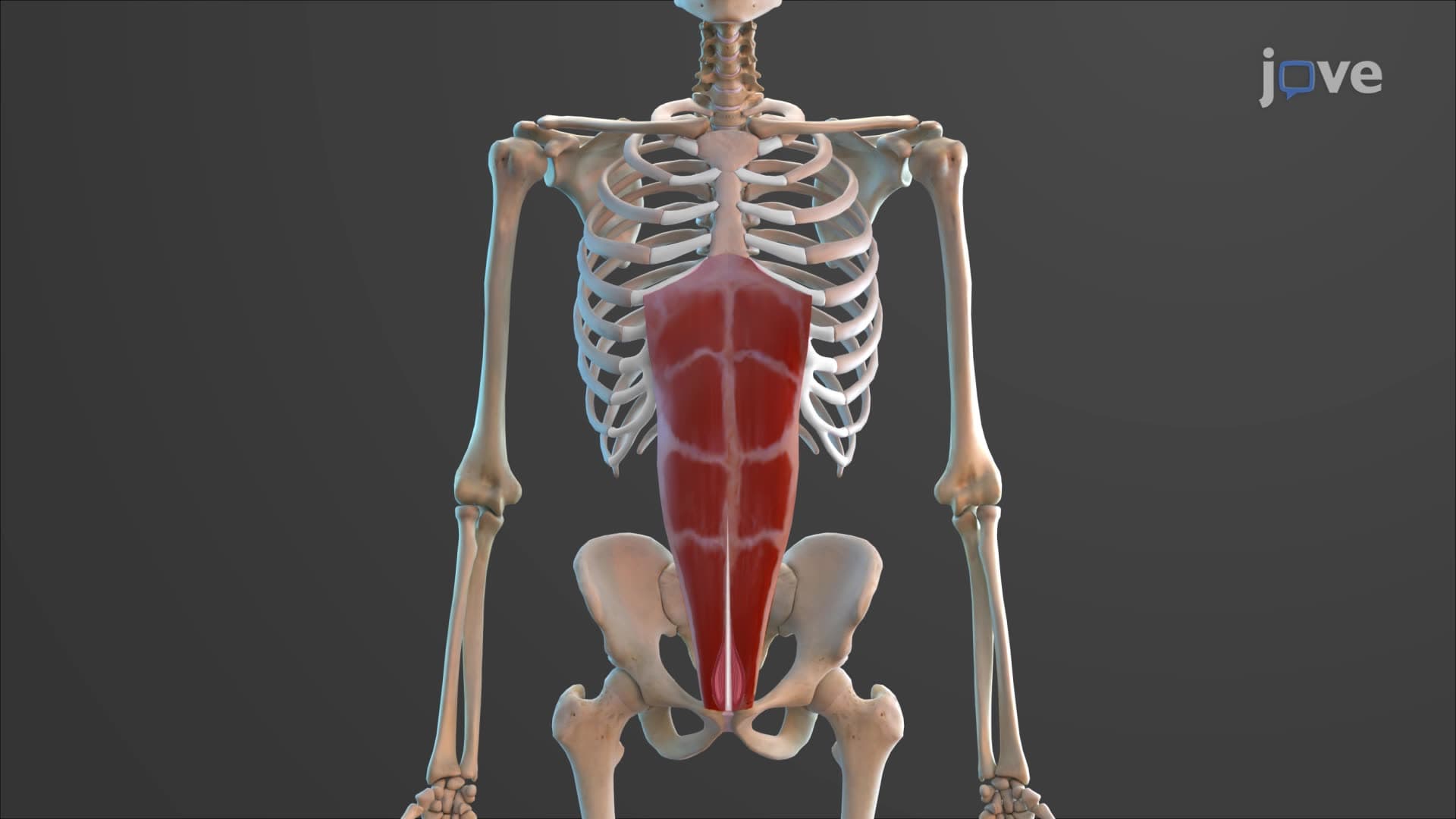
12. Muscles of the Abdomen
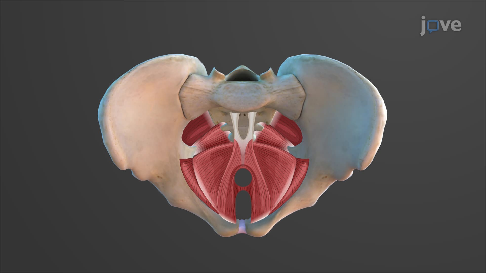
13. Muscles of the Pelvic Floor and Perineum
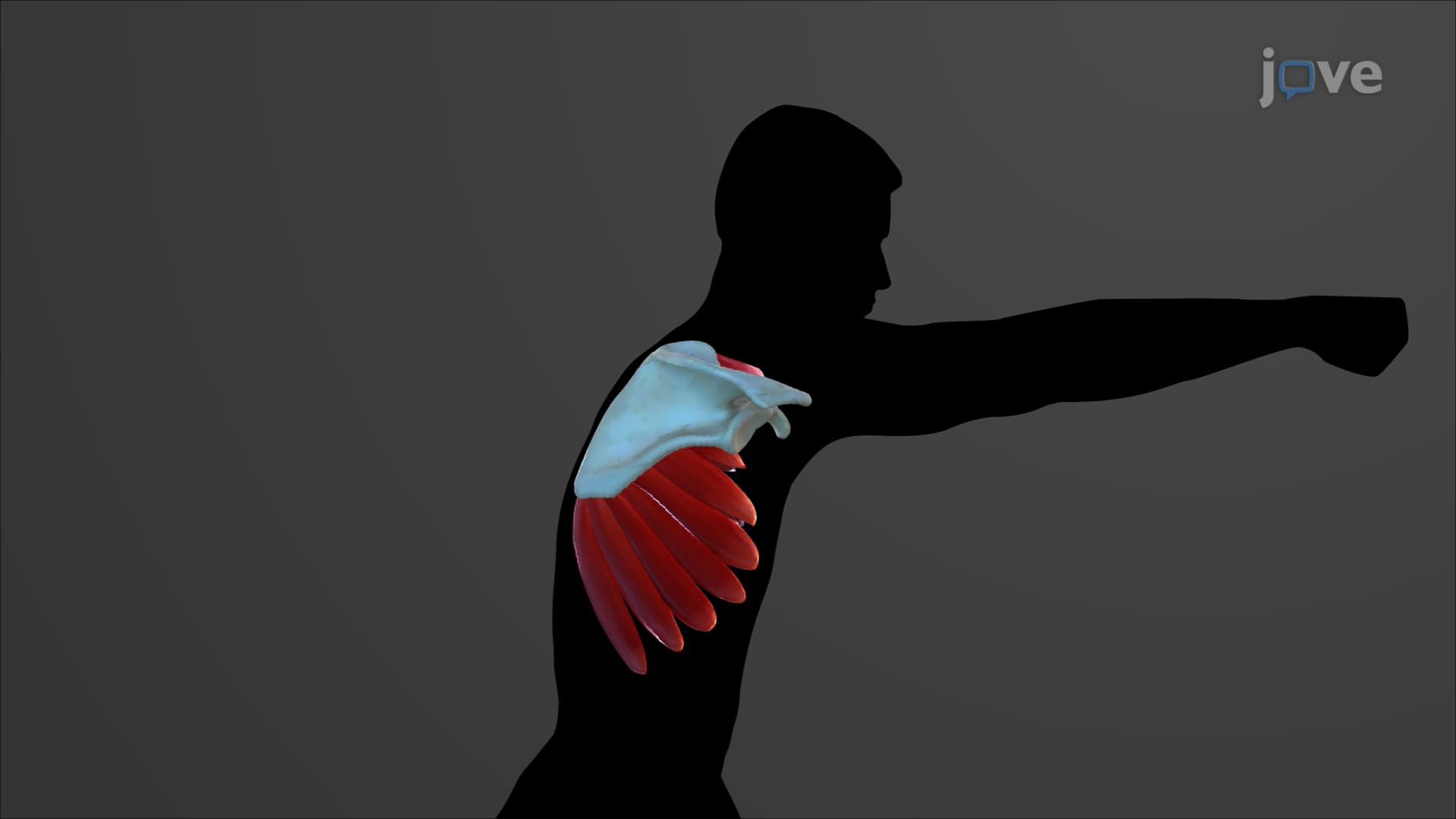
14. Muscles of the Shoulder
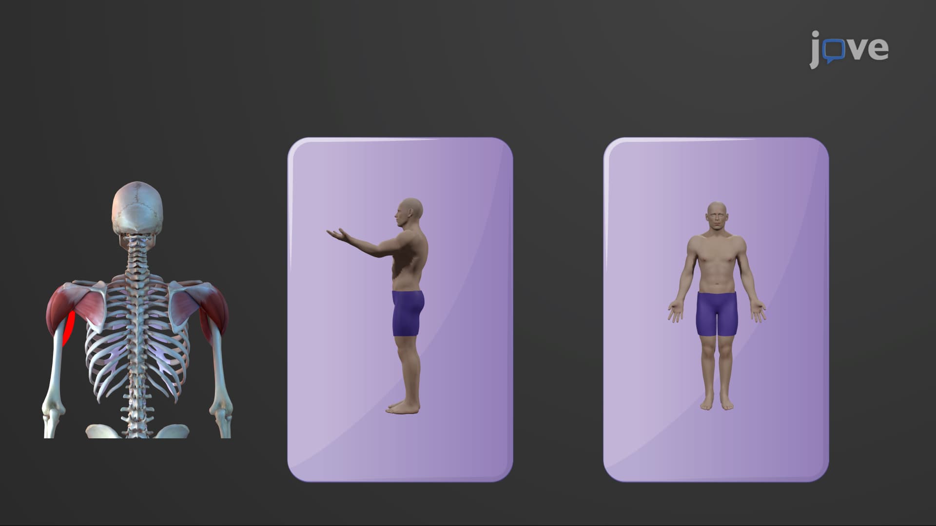
15. Muscles that Move the Arm
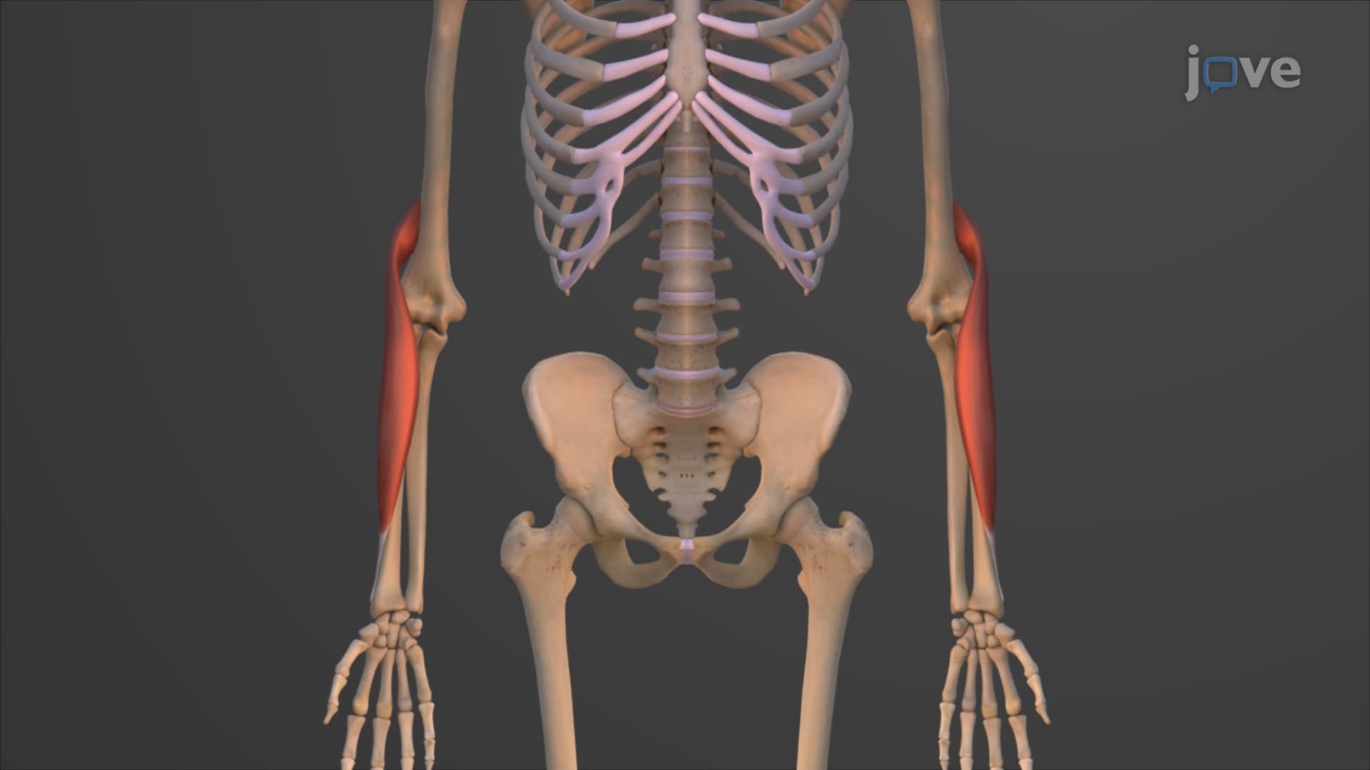
16. Muscles that Move the Forearm
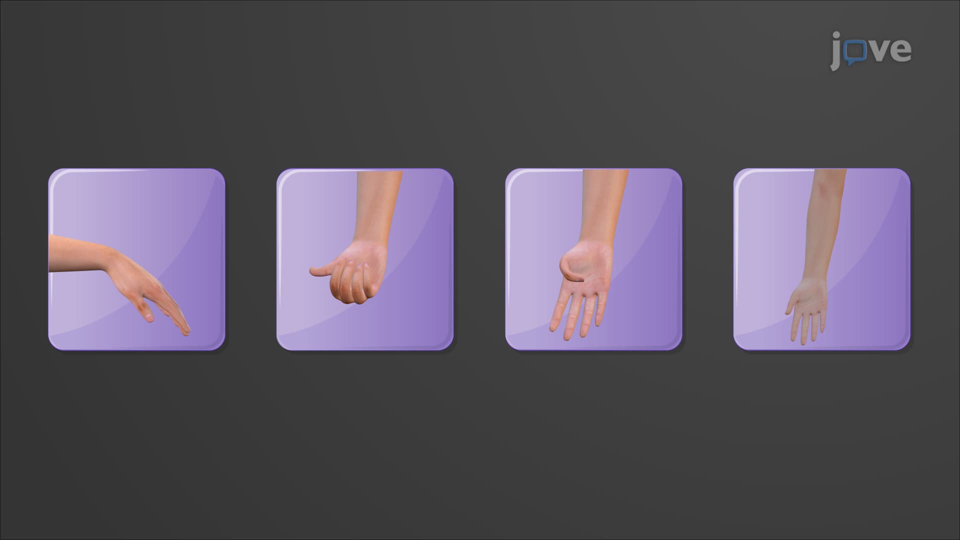
17. Muscles of the Forearm that Move the Hand and Fingers
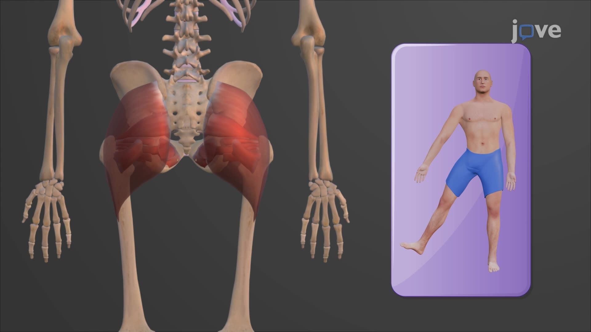
18. Muscles that Move the Thigh
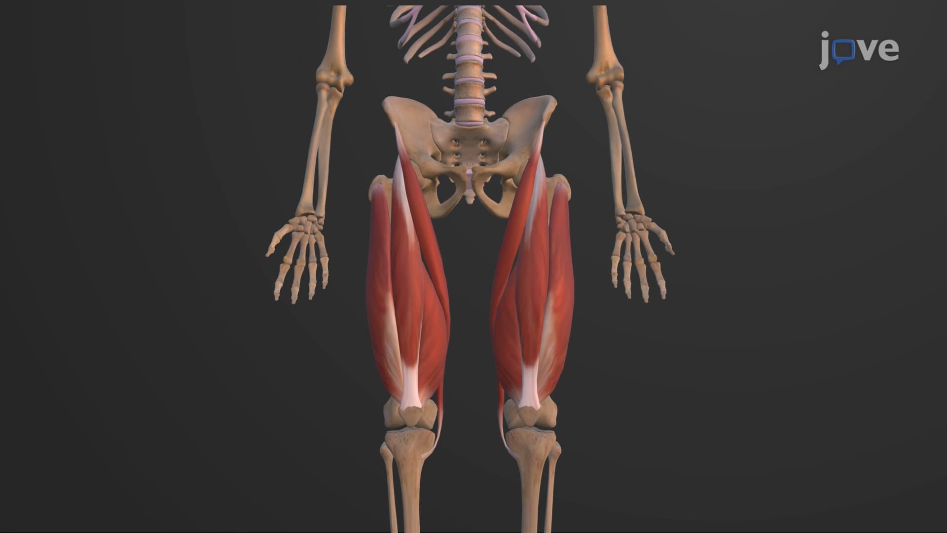
19. Muscles that Move the Leg
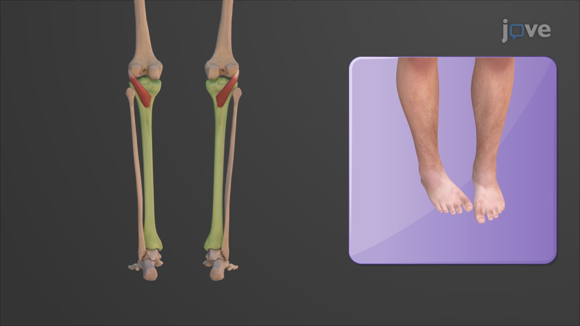
20. Muscles of the Leg that Move the Foot and Toes
The muscular system forms the foundation of human movement and body function, comprising skeletal, smooth, and cardiac muscles that work together to enable everything from voluntary motion to involuntary processes like digestion and circulation. This comprehensive course explores human muscle anatomy through detailed examination of muscle structure, fascicle arrangements, coordination mechanisms, and the major muscles and their functions in the body across axial and appendicular regions. Using JoVE Coach's systematic approach, students will master essential concepts for success in AP Biology, anatomy courses, and pre-health programs.
- Understand the three types of muscle tissue and their distinct roles in the muscular system
- Identify fascicle arrangements and how they determine muscle function and force generation
- Analyze muscle coordination patterns including agonist, antagonist, synergist, and fixator relationships
- Learn systematic approaches to naming skeletal muscles based on location, shape, and function
- Explore the organization of axial muscles controlling head, neck, trunk, and respiratory movements
- Examine appendicular muscles that control limb movement and joint stability
- Apply knowledge of lever systems and neuromuscular junction principles to understand muscle mechanics
- Understand motor unit recruitment and how the nervous system controls muscle contraction
1. Muscle Tissue Classification and Structure: The three primary muscle types—skeletal, smooth, and cardiac—each serve distinct physiological roles. Skeletal muscles form the musculoskeletal system with bones and connective tissues, enabling voluntary movement through contractions that move articulating bones. Each skeletal muscle contains origins (proximal attachments to stationary bones), insertions (distal attachments to movable bones), and bellies (fleshy central regions). Understanding this basic architecture explains how muscles like the biceps brachii generate force to flex the elbow, with origins on the scapula serving as stable anchor points while the insertion on the radius creates movement. This structural organization underlies all voluntary motor function in the human body.
2. Fascicle Arrangements and Muscle Architecture: Skeletal muscles are organized into fascicles—bundles of muscle fibers surrounded by perimysium connective tissue—arranged in four distinct patterns that determine functional capacity. Parallel muscles like the sartorius have fascicles aligned with the muscle's long axis, maximizing range of motion. Convergent muscles such as the pectoralis major converge widespread fascicles to a single attachment point, combining power with versatility. Pennate muscles (unipennate, bipennate, multipennate) attach fascicles obliquely to tendons, maximizing force production at the expense of range. Circular muscles like the orbicularis oris form sphincters around body openings. These architectural patterns directly correlate with functional demands, explaining why pennate muscles excel at generating force while parallel muscles provide greater movement range.
3. Muscle Coordination and Movement Mechanics: Coordinated movement requires precise interaction between muscle groups functioning as agonists, antagonists, synergists, and fixators. Agonist muscles like the biceps brachii during elbow flexion produce primary movement, while antagonists such as the triceps brachii provide opposing force for control and reversal. Synergist muscles assist agonists by stabilizing joints and adding force, exemplified by the brachialis supporting biceps function. Fixator muscles, including the rotator cuff group, stabilize proximal joints to enable distal movement efficiency. This coordination system operates through complex neuromuscular junction interactions where motor units recruit muscle fibers based on force requirements, creating smooth, controlled movements essential for activities from writing to weightlifting.
4. Systematic Muscle Nomenclature: Anatomical muscle names follow logical patterns based on location, physical characteristics, attachments, and function, creating a systematic approach to learning hundreds of individual muscles. Location-based names like "brachii" (of the arm) immediately indicate anatomical position, while descriptive terms such as "deltoid" (triangular) describe shape characteristics. Attachment-based names list origin before insertion, as seen in the stylohyoid muscle connecting the temporal bone's styloid process to the hyoid bone. Functional names incorporate action words like "adductor" or "extensor," while size indicators such as "longus," "brevis," "major," and "minor" distinguish between related muscles. This nomenclature system transforms muscle memorization from rote learning into logical pattern recognition.
5. Axial Muscle Organization and Function: Axial muscles along the body's midline control essential functions including facial expression, respiration, spinal movement, and core stability. Facial expression muscles like the occipitofrontalis and orbicularis oris originate from facial bones and insert into skin, creating diverse expressions through coordinated contractions. Respiratory muscles including the diaphragm and intercostals generate breathing movements through coordinated thoracic cavity volume changes. Vertebral column muscles organized in superficial, intermediate, and deep layers provide spinal stability and movement, with erector spinae muscles enabling extension and lateral flexion. Abdominal muscles form the anterolateral trunk wall, creating the linea alba through interwoven aponeuroses while providing core stability essential for all movement patterns and protecting internal organs.
6. Appendicular Muscle Systems: Appendicular muscles associated with limb girdles and extremities enable complex movements from fine motor control to powerful locomotion. Upper limb muscles progress from shoulder stabilizers like the rotator cuff through arm movers including the deltoid and pectoralis major, to forearm controllers such as the biceps and triceps, finally reaching intricate hand and finger muscles enabling precision grip. Lower limb muscles follow similar organization with hip stabilizers like the gluteal complex, thigh movers including the quadriceps and hamstrings, and foot controllers in the lower leg compartments. This hierarchical organization allows for both gross motor movements like running and fine motor control like surgical procedures, with each muscle group contributing specific actions within coordinated movement patterns essential for human locomotion and manipulation.
Frequently Asked Questions
Agonist muscles produce the primary movement at a joint, while antagonist muscles create the opposite action and are typically located on the opposite side of the bone. For example, during elbow flexion, the biceps brachii acts as the agonist by contracting to bend the arm, while the triceps brachii serves as the antagonist. We need both because antagonists provide controlled resistance during movement (preventing jerky motions), enable smooth deceleration, and reverse the movement when needed. This partnership creates the precise, controlled movements essential for everything from typing to throwing a baseball.
Focus on understanding the naming patterns rather than pure memorization. Group muscles by location (all arm muscles together), then learn the systematic naming conventions: location terms (brachii = arm, femoris = thigh), shape descriptors (deltoid = triangular), size indicators (major/minor, longus/brevis), and function words (flexor, extensor, adductor). Create anatomical diagrams with muscle groups color-coded by function, and practice identifying muscles on skeletal models. The College Board emphasizes understanding muscle coordination and function over memorizing every individual muscle name.
The MCAT focuses on functional relationships rather than detailed anatomy. Prioritize understanding muscle tissue types, the sliding filament mechanism, neuromuscular junction physiology, and how muscles work as lever systems with bones and joints. Know major muscle groups and their primary functions (quadriceps extend the knee, hamstrings flex it), muscle coordination patterns (agonist/antagonist relationships), and how muscle architecture affects force and range of motion. Understand respiratory muscle mechanics and how core muscles provide stability during movement, as these concepts frequently appear in passage-based questions.
Fascicle arrangement directly determines muscle performance characteristics. Parallel muscles like the sartorius maximize range of motion because fascicles run the full muscle length, but they generate less force. Pennate muscles like the deltoid pack more fascicles into the same space by angling them relative to the tendon, creating greater force production but reduced range of motion. Convergent muscles like the pectoralis major combine benefits by spreading fascicles over a wide origin area then converging to a single insertion, providing both power and versatility. Circular muscles form sphincters that can completely close openings, like the orbicularis oris around the mouth.
The posterior muscles are challenging because they're organized in three overlapping layers (superficial, intermediate, deep) with similar names and functions. Start with the big picture: superficial muscles like the trapezius move the head and shoulder blades, intermediate erector spinae muscles extend the spine, and deep muscles fine-tune individual vertebrae positions. Focus on function first—muscles that extend the spine, rotate it, or provide lateral flexion—then learn the specific names. Use the anatomical position principle: muscles closer to the midline handle spinal movement, while those toward the sides manage head and shoulder blade actions.
Breathing involves coordinated action between the diaphragm and intercostal muscles. During inspiration, the diaphragm contracts and flattens, increasing vertical thoracic cavity space, while external intercostal muscles contract to elevate ribs, expanding the cavity laterally and anteriorly. This creates negative pressure that draws air into the lungs. During expiration, the diaphragm relaxes and internal intercostal muscles contract to pull ribs together, decreasing cavity volume and pushing air out. The accessory muscles of respiration (neck and abdominal muscles) assist during forced breathing, such as during exercise or respiratory distress.
Connect muscle functions to real-world movements you perform daily. When studying arm muscles, actually perform the motions—flex your biceps, extend with your triceps, rotate your forearm. For leg muscles, walk up stairs (quadriceps extend the knee), walk downhill (hamstrings control knee flexion), and stand on your toes (calf muscles plantar flex). Create movement scenarios: "throwing a baseball uses shoulder muscles for arm movement, core muscles for stability, and leg muscles for power transfer." This functional approach helps you understand not just individual muscle actions, but how muscle groups coordinate to produce complex movements essential in sports, physical therapy, and daily activities.
This microcourse includes 20 concept videos that walk you through the building blocks of Anatomy and Physiology. Each video is short, about 1 minute, so you can cover a full topic during a coffee break or between classes. The full sequence starts with The Muscular System and ends with Muscles of the Leg that Move the Foot and Toes.
The playlist moves from big-picture ideas to the precise vocabulary used in Anatomy and Physiology. Early videos introduce The Muscular System, Fascicle Arrangement in Skeletal Muscles, and Muscle Coordination and Action. The middle of the series focuses on Axial and Appendicular Muscles, Muscles for Facial Expressions, and Muscles of the Eye. The final stretch covers Muscles of the Anterior Neck, Muscles that Move the Head, Muscles of the Vertebral Column, Muscles of the Thorax, Muscles of the Abdomen, Muscles of the Pelvic Floor and Perineum, and Muscles of the Leg that Move the Foot and Toes.
The natural next step is The Nervous System and Nervous Tissue. From there, you can move to Anatomy of the Central and Peripheral Nervous System, Functions of the Central and Peripheral Nervous System, and The Autonomic Nervous System. Once you finish those, the full Anatomy and Physiology curriculum of 31 microcourses on JoVE Coach opens up, taking you from foundational concepts to advanced systems.
Related Subjects
 Medical-Surgical Nursing 28 micro-courses›
 Nursing 14 micro-courses›