Loading video…

 12 Concepts
 2.3K views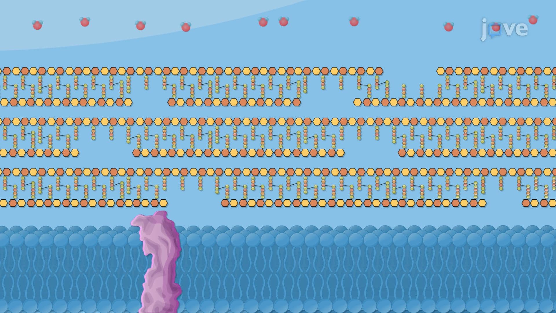

Video Summary: Inhibitors of Gram Positive Cell Wall Synthesis Explained
Did you know that a single antibiotic can destroy billions of bacteria by targeting the very wall that keeps them alive? Inhibitors of gram-positive cell wall synthesis work by disrupting peptidoglycan, the structural backbone of bacterial cells. Penicillin, one of the most prescribed antibiotics in US hospitals, exemplifies this mechanism by blocking proteins that stitch bacterial walls together. Watch the full video on JoVE Coach to master this concept with expert-led visuals and step-by-step explanations.
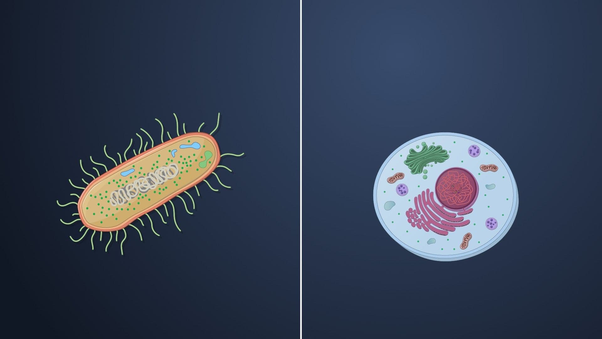
Bacteria are fundamentally different from human cells in one critical way: they build rigid cell walls made of peptidoglycan, a mesh-like polymer that human cells do not produce. This makes peptidoglycan an ideal target for antibiotics, drugs can attack it without directly harming the patient's own cells. Understanding inhibitors of gram-positive cell wall synthesis is foundational to microbiology, pharmacology, and clinical medicine, and it appears regularly on AP Biology exams, college microbiology midterms, and high-stakes tests like the MCAT and USMLE Step 1.
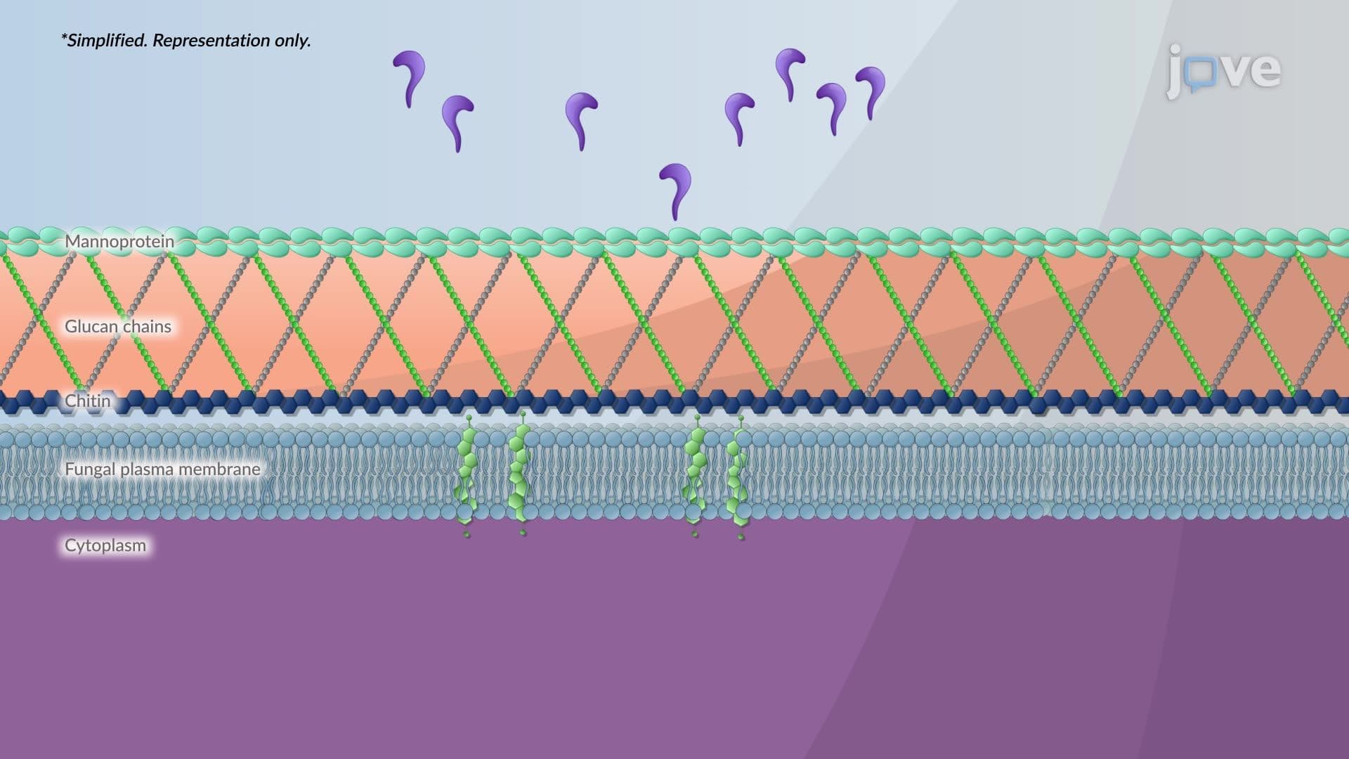
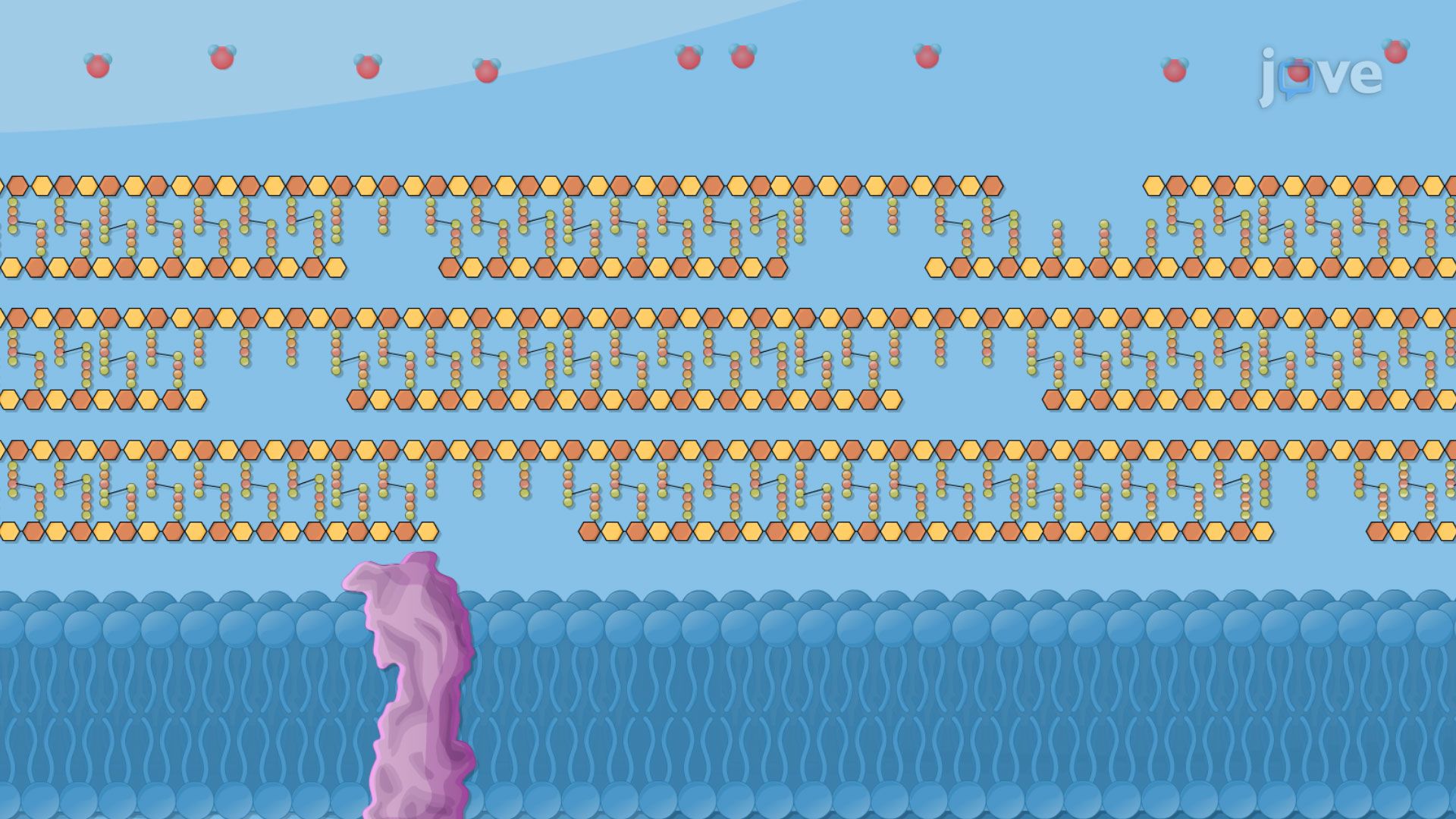
Peptidoglycan is a unique macromolecule found in bacterial cell walls. It consists of long sugar chains, made of alternating N-acetylglucosamine (NAG) and N-acetylmuramic acid (NAM), cross-linked by short peptide bridges. This lattice-like structure functions like a molecular cage, protecting the bacterium from bursting under osmotic pressure. Gram-positive bacteria, such as *Staphylococcus aureus* and *Streptococcus pyogenes* (common causes of US emergency room infections), have an especially thick peptidoglycan layer, making them both robust and uniquely vulnerable to drugs that disrupt this layer.
Beta-lactam antibiotics, which include penicillins, cephalosporins, and carbapenems, share a defining structural feature: the four-membered beta-lactam ring. This ring closely resembles the natural bacterial substrate D-alanyl-D-alanine, a dipeptide that penicillin-binding proteins (PBPs) normally recognize and process during the cross-linking phase of cell wall synthesis.
When a beta-lactam antibiotic enters a bacterial cell, its ring covalently bonds to the active site of a PBP, essentially jamming the enzyme permanently. With PBPs inactivated, bacteria can no longer stitch their glycan chains together. The cell wall becomes fragile and porous. Simultaneously, naturally occurring autolytic enzymes called murein hydrolases continue breaking down existing peptidoglycan. The result is catastrophic structural failure, the bacterial membrane ruptures under osmotic pressure, and the cell dies. This makes beta-lactams bactericidal drugs, meaning they actively kill bacteria rather than simply slowing their growth.
Not all beta-lactams work equally across all bacteria. Penicillin G, for example, is considered a narrow-spectrum antibiotic, highly effective against gram-positive organisms but limited against gram-negative bacteria, which have an outer membrane that restricts drug entry. In contrast, broad-spectrum antibiotics like ampicillin can target a wider range of organisms. In US clinical practice, choosing the right drug requires identifying the pathogen first, a process called culture and sensitivity testing, to avoid unnecessary broad-spectrum use that can drive antimicrobial resistance.
One of the most urgent challenges in modern US healthcare is antimicrobial resistance, particularly against beta-lactam drugs. Methicillin-resistant *Staphylococcus aureus* (MRSA), a major concern in hospitals across the country, produces an altered PBP called PBP2a, encoded by the *mecA* gene. This modified protein has low affinity for beta-lactam antibiotics, meaning the drugs can no longer bind effectively. Understanding how bacteria develop resistance to antibiotics through mechanisms like PBP alteration, beta-lactamase enzyme production, and efflux pumps is essential for anyone studying infectious disease, pharmacology, or preparing for exams like the NCLEX or USMLE.
Related Micro-courses