Loading video…

 14 Concepts
 2.1K views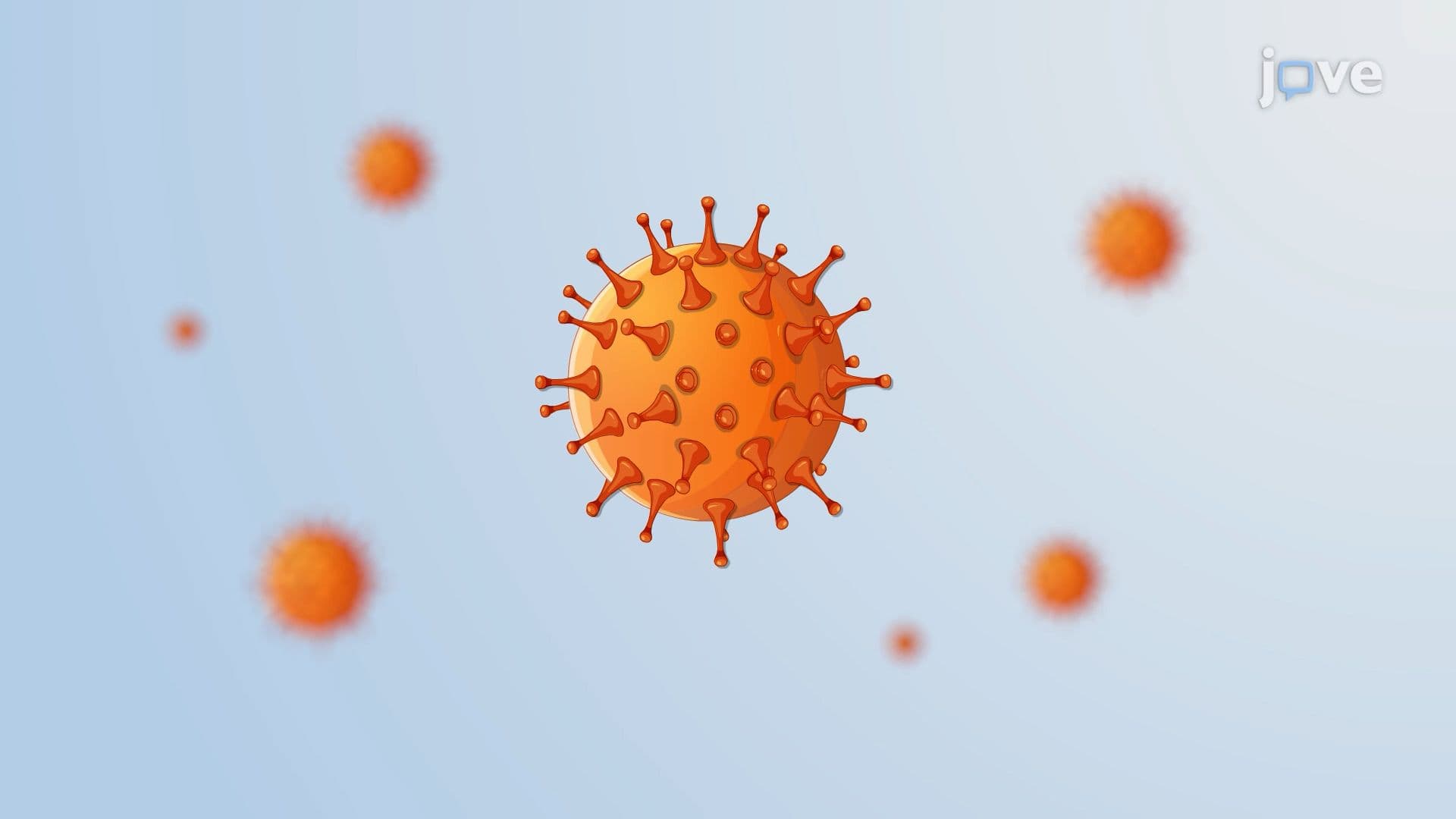

Video Summary: What Is Cytomegalovirus Disease
Did you know a virus can hide inside your immune cells for years, then strike when your defenses drop? Cytomegalovirus disease, caused by a stealthy herpesvirus, affects millions of Americans, especially organ transplant recipients whose immunity is suppressed. This pathogen evades detection by blocking the body's ability to flag infected cells for destruction. Watch the full video on JoVE Coach to master this concept with expert-led visuals and step-by-step explanations.
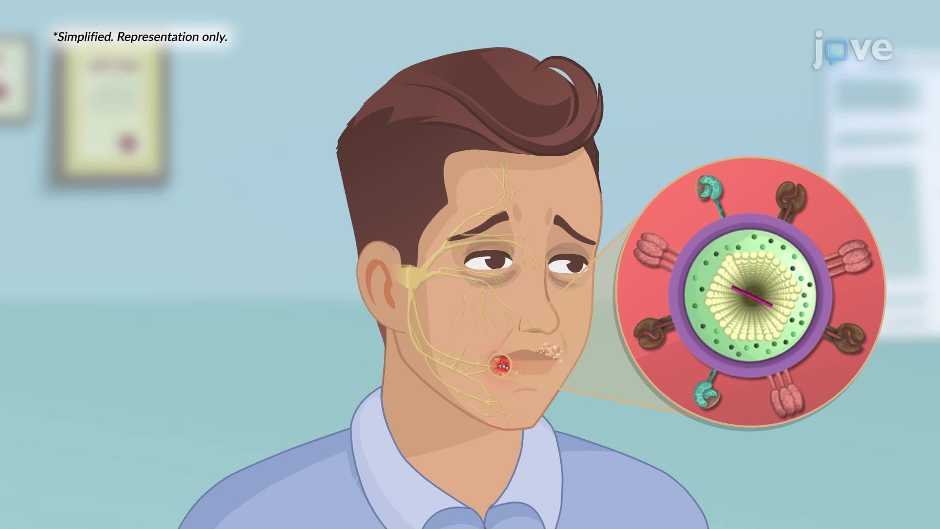
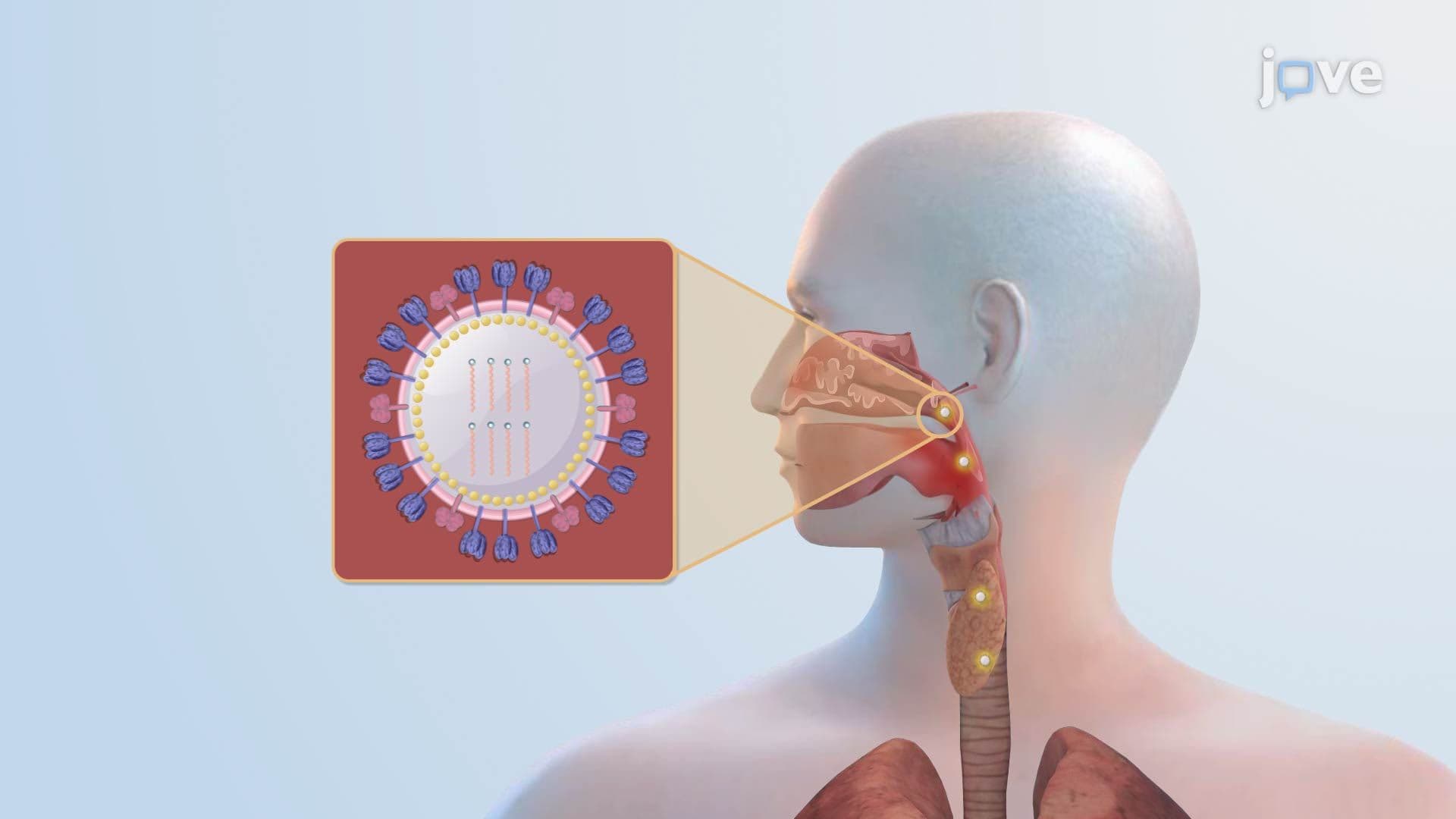
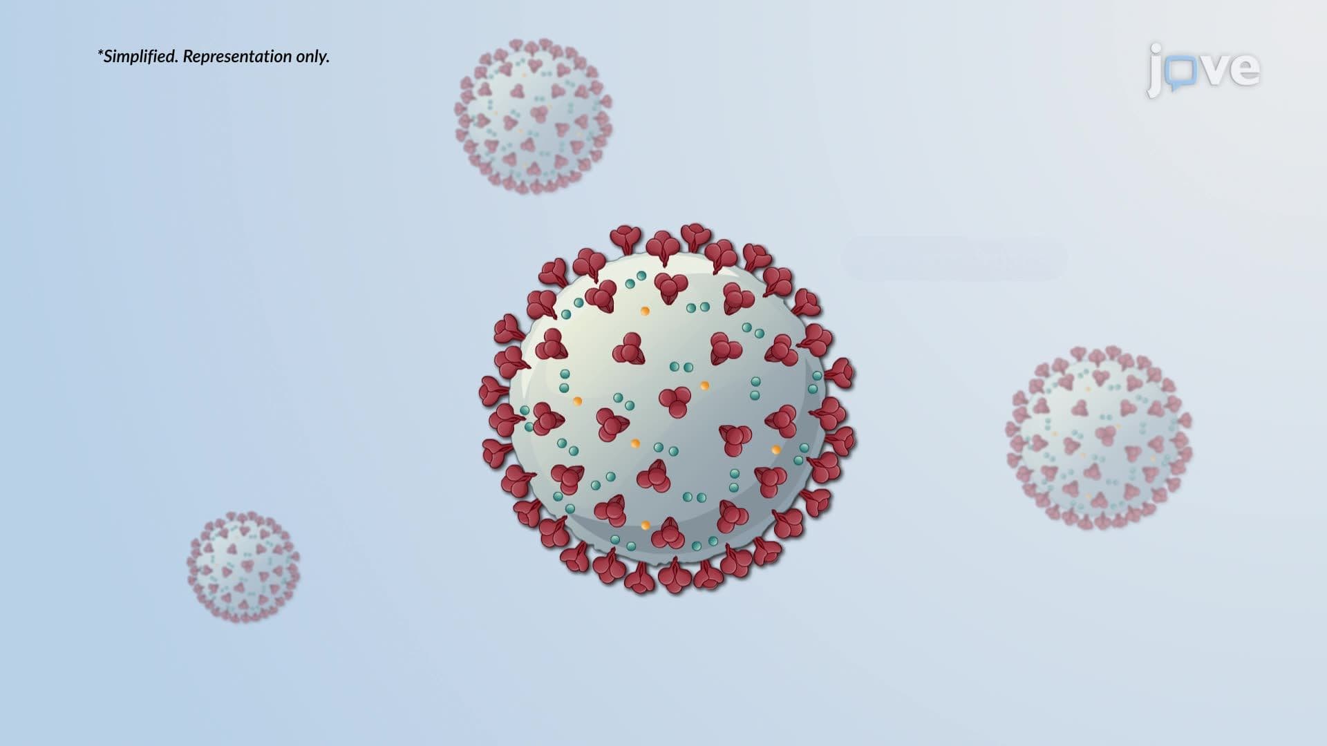
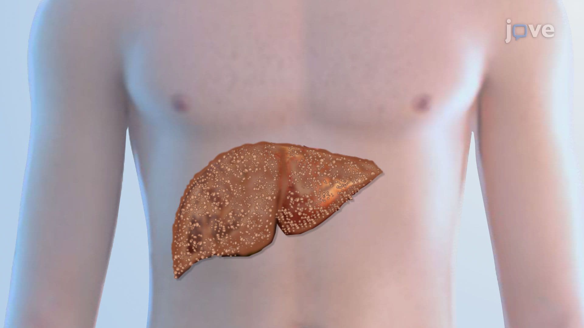
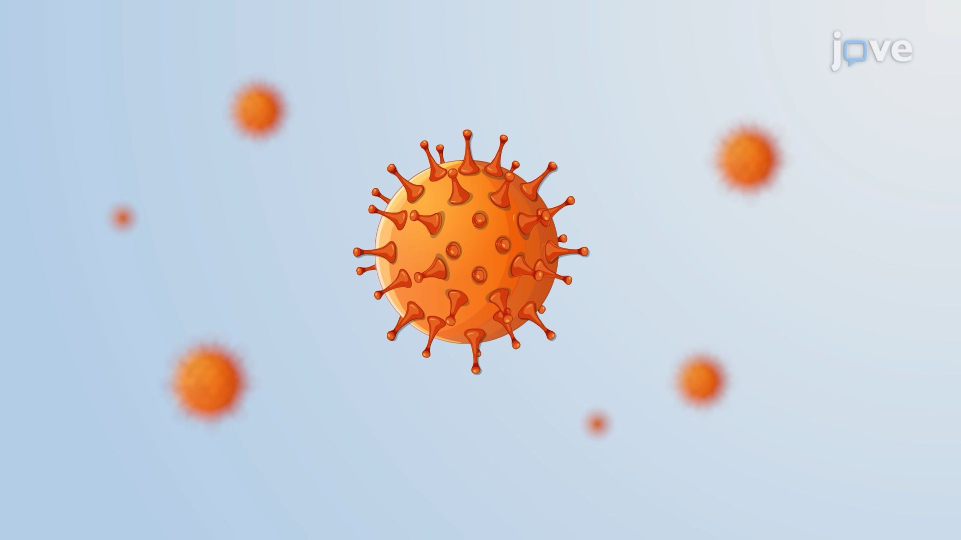
Cytomegalovirus disease is caused by Human Cytomegalovirus (HCMV), a double-stranded DNA virus belonging to the *Herpesviridae* family. It is one of the most clinically significant viral pathogens in immunocompromised patients in the United States. While CMV infects an estimated 50-80% of American adults by age 40 with few or no symptoms in healthy individuals, it becomes life-threatening when immunity is suppressed, making it a high-priority concern in organ transplant medicine, HIV/AIDS care, and neonatal health.
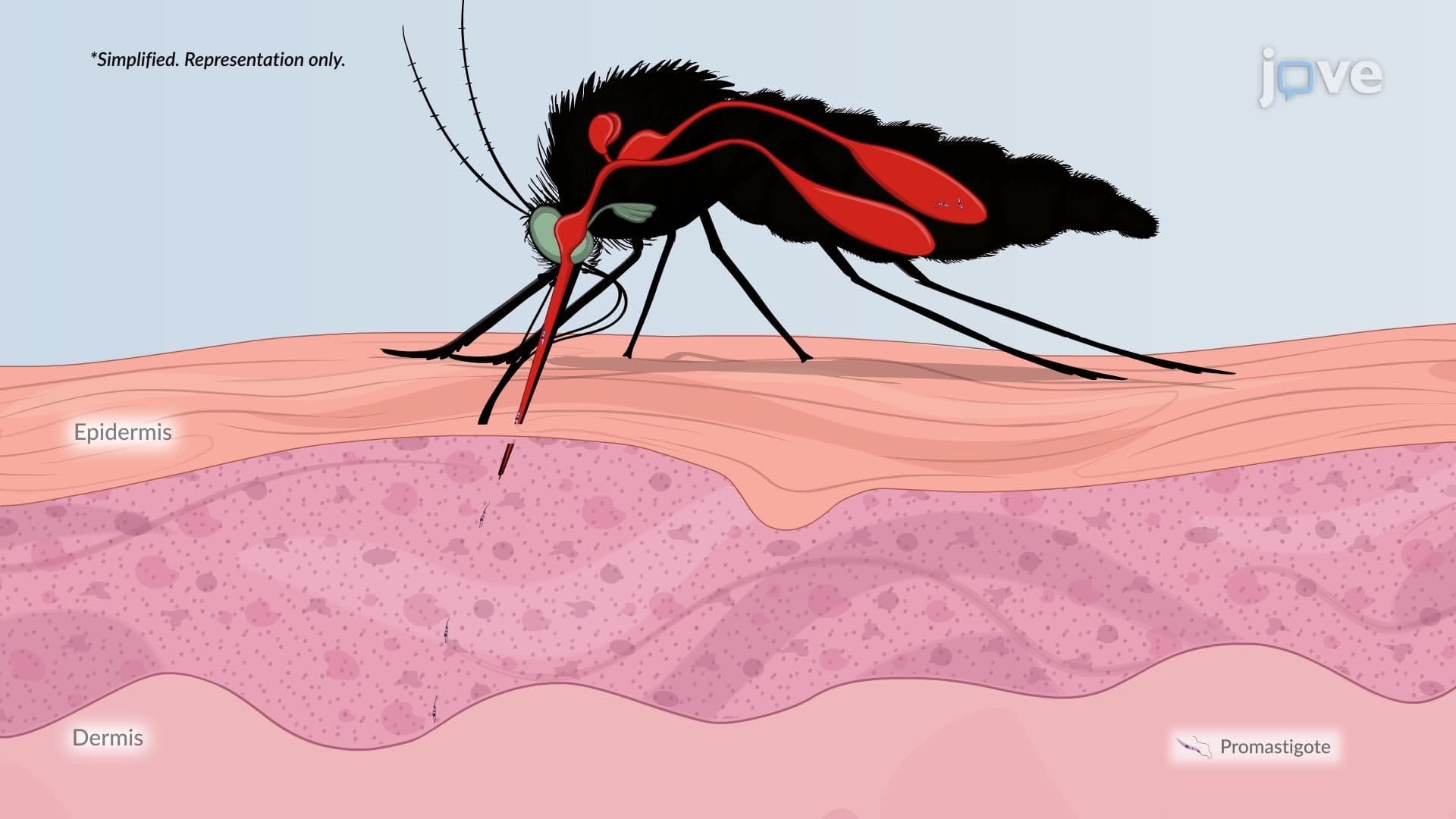
CMV spreads through contact with infected body fluids. In clinical settings across the US, transmission occurs during solid organ or bone marrow transplantation, blood transfusions, and from mother to fetus, a process called vertical transmission. Congenital CMV is the leading infectious cause of birth defects in the United States, affecting approximately 1 in 200 newborns annually. Sexual transmission is also documented. Understanding these routes is essential for AP Biology students studying viral pathogenesis and for pre-med undergraduates preparing for the MCAT, where host-pathogen interactions are frequently tested.
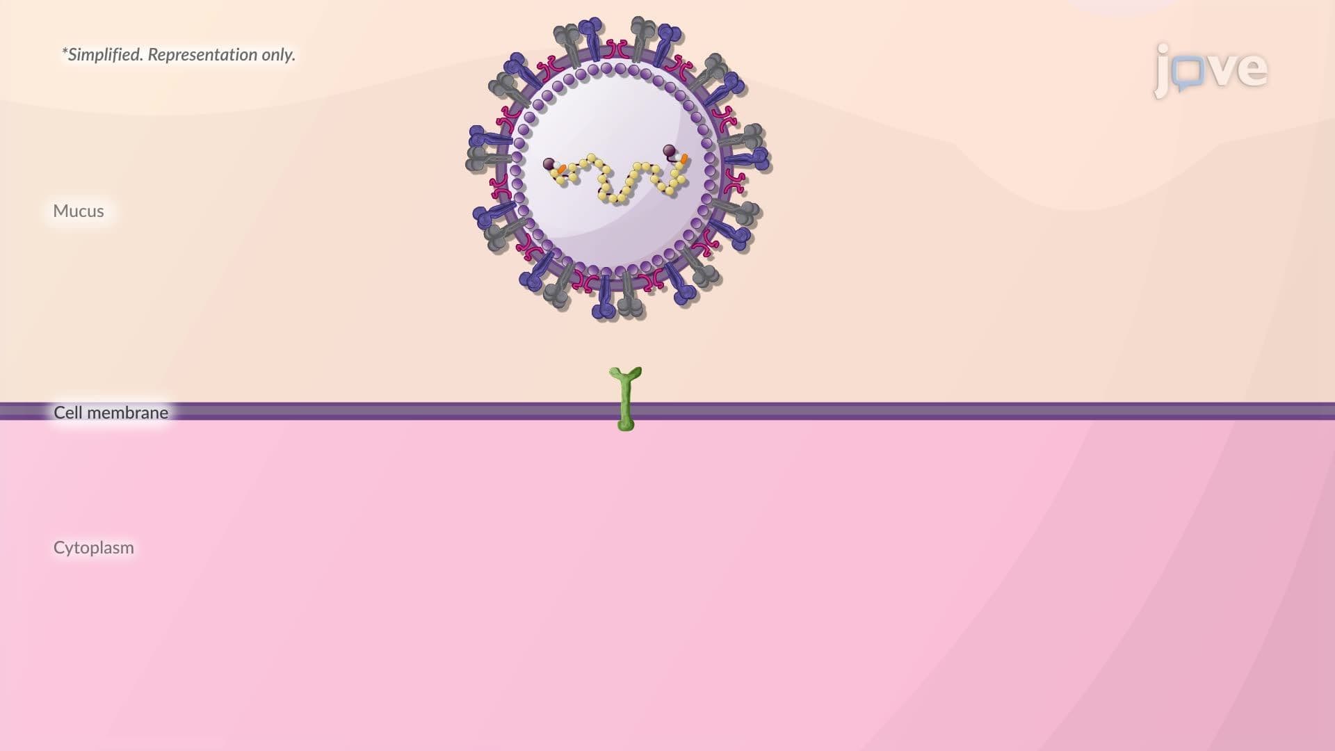
One of the most remarkable features of CMV, and of herpesviruses generally, is their ability to establish latency. After initial infection, CMV embeds itself in monocytes and hematopoietic progenitor cells (the precursor cells that give rise to blood cells in bone marrow), essentially going dormant. The virus is not eliminated; it simply pauses replication. When a patient's immune system is weakened, for example, after receiving immunosuppressive drugs following a kidney or heart transplant at a hospital like Mayo Clinic or Johns Hopkins, the virus reactivates, hijacks the host cell's replication machinery, and produces new viral particles called virions that spread the infection.
This is where Cytomegalovirus disease gets especially fascinating from a virology standpoint. CMV actively dismantles two key immune defense systems:
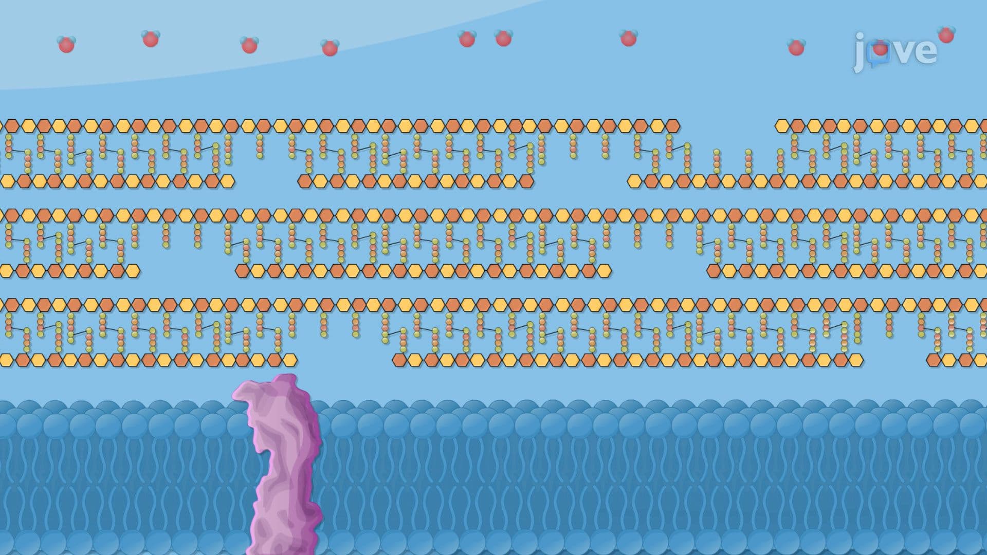
1. MHC Class I Interference: Normally, infected cells display viral protein fragments on MHC class I molecules, flagging themselves for destruction by CD8+ cytotoxic T cells. CMV encodes proteins that block this antigen presentation pathway, essentially making infected cells "invisible" to the immune system's hit squad.
2. Chemokine Disruption: Certain CMV proteins bind to host chemokines, chemical signals that recruit immune cells to the infection site, effectively jamming the body's alarm system and preventing an adequate immune response from assembling.
These evasion strategies are a cornerstone topic in college-level microbiology and immunology courses, and they appear in USMLE Step 1 review materials under immune evasion mechanisms. For AP Biology students, they illustrate how viruses evolve sophisticated molecular strategies to exploit host biology.
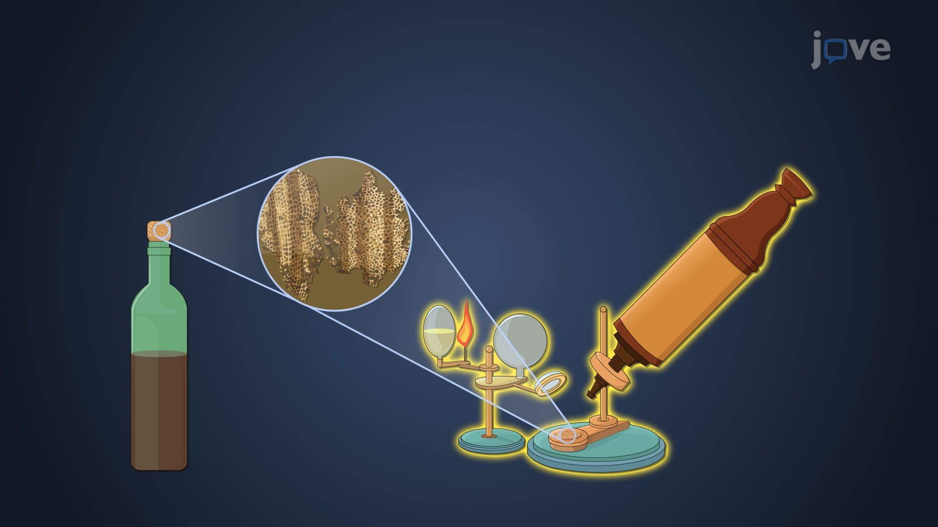
Under a microscope, cells infected with CMV undergo a distinctive transformation: they become dramatically enlarged (a phenomenon called cytomegaly, which literally gives the virus its name) and develop intranuclear inclusion bodies, large, dark-staining masses inside the cell nucleus surrounded by a clear halo. This "owl's eye" appearance is a classic pathology finding tested on the USMLE, NCLEX, and college pathology exams. Recognizing this microscopic signature connects the visual world of histology to the molecular biology of viral replication, a bridge that reinforces learning across multiple science disciplines.
Related Micro-courses